Post-cataract corneal edema is stromal or epithelial swelling that appears after lens surgery, usually within the first days.
Most cases are mild and clear on their own.
However, some eyes do not recover, and the timeline shows which ones need action.
What Causes Post-Cataract Corneal Edema
The endothelial pump keeps the stroma relatively dehydrated.
During phacoemulsification, ultrasound energy, turbulence and instrument contact injure endothelial cells.
As a result, pump function drops and fluid enters the stroma.
Because adult endothelial cells barely divide, neighboring cells must enlarge and spread to cover the gap.
How Long Post-Cataract Corneal Edema Lasts
Mild edema near the main incision is common on day one.
In routine cases, it usually fades within a few days.
Moderate diffuse edema often clears over one to several weeks.
Therefore, a hazy cornea on day one is rarely an emergency by itself.
All-fit Slit-Lamp Adapter
Record and share exactly what you see at the slit lamp. One adapter fits any slit lamp or surgical microscope — and any smartphone.
From Choroida — the team behind this siteStriate Keratopathy
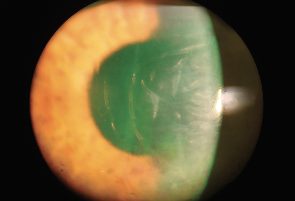
Striate keratopathy appears as fine gray lines in the posterior stroma.
These lines are folds in Descemet membrane caused by stromal swelling.
They are common after a dense nucleus or long surgery, and they usually fade as the endothelium recovers.
What the Patient Reports
Patients describe blur or a foggy view that is often worse on waking.
Because the lids are closed overnight, less water evaporates and the cornea stays thicker in the morning.
Vision then improves through the day.
Examining Post-Cataract Corneal Edema
First, map the edema as localized, sectoral or diffuse.
Next, check the wound, the anterior chamber reaction and the intraocular pressure.
Then inspect the inferior angle for a retained fragment, using gonioscopy when the cornea allows.
Finally, look for guttae in the fellow eye, which suggest Fuchs dystrophy (see causes of corneal edema).
Pressure Spikes in Post-Cataract Corneal Edema
Retained viscoelastic can raise intraocular pressure within hours of surgery.
High pressure pushes fluid into the epithelium and produces microcystic edema.
Therefore, lowering the pressure often clears the cornea quickly.
Localized Edema and Mechanical Causes
Persistent sectoral edema points to a structural problem.
A detachment near the incision is a classic cause (see Descemet membrane detachment).
Anterior segment OCT confirms it and guides air or gas tamponade.
Also, a nuclear fragment in the inferior angle can sustain inferior edema until removal.
TASS Versus Infection
Toxic anterior segment syndrome (TASS) is a sterile reaction to material entering the eye during surgery.
It typically starts within 12 to 24 hours with diffuse limbus-to-limbus edema.
In contrast, endophthalmitis usually appears later, with pain, vitritis and progressive inflammation.
Because the two can overlap, any doubt warrants urgent evaluation for infection.
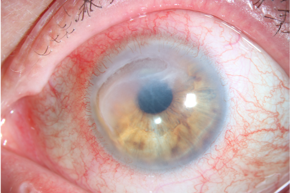
When to Suspect Decompensation
Edema that worsens after the first weeks is a warning sign.
Similarly, a cornea that stays thick and hazy at about three months is unlikely to clear fully.
Epithelial bullae and recurrent pain then suggest pseudophakic bullous keratopathy (see endothelial decompensation).
Tests That Help
Serial pachymetry shows whether the cornea is thinning toward normal.
Specular microscopy is often unreadable while edema persists.
However, the fellow eye offers a useful estimate of endothelial reserve.
Medical Treatment of Post-Cataract Corneal Edema
Topical corticosteroids control postoperative inflammation, which can delay endothelial recovery.
Hypertonic sodium chloride 5% drops or ointment draw water from the epithelium.
They ease morning blur, although they do not restore pump function.
In addition, use topical carbonic anhydrase inhibitors cautiously when the endothelium is compromised.
Rho Kinase Inhibitors
Ripasudil and netarsudil have been studied as agents to speed endothelial recovery.
Some early reports describe faster clearing after surgery.
However, the evidence is limited, and this use remains off-label.
Surgical Options
When edema persists, endothelial keratoplasty is the usual next step.
DMEK and DSAEK replace the failed endothelium with a thin donor layer.
Meanwhile, a bandage contact lens can relieve pain from ruptured bullae.
Counseling About Post-Cataract Corneal Edema
Tell patients that early cloudiness is common and usually temporary.
Explain that vision may fluctuate for several weeks.
Then give clear return precautions: increasing pain, redness or a sudden drop in vision.
For eyes with Fuchs dystrophy, discuss the chance of later keratoplasty before surgery.
Preventing Post-Cataract Corneal Edema
Preoperative pachymetry and guttae assessment identify high-risk eyes.
Dispersive viscoelastic also protects the endothelium during long phaco.
In addition, lower energy and phaco away from the cornea reduce cell loss.
Finally, thorough viscoelastic removal limits pressure spikes.
Pitfalls to Avoid
- Do not label every day-one haze as TASS or infection
- Check localized edema early for a Descemet detachment
- Never miss an inferior retained fragment
- Do not taper steroids too fast while the cornea is swollen
- Avoid predicting final vision before endothelial reserve is known


Document what you see
Two smartphone imaging tools built for everyday clinic use — one for the slit lamp, one for the fundus.
From Choroida — the team behind this siteReferences
- Salmon JF. Kanski’s Clinical Ophthalmology: A Systematic Approach. 9th ed. Elsevier; 2020.
- American Academy of Ophthalmology. Basic and Clinical Science Course, Section 11: Lens and Cataract. San Francisco: AAO; 2023.
- Mamalis N, Edelhauser HF, Dawson DG, et al. Toxic anterior segment syndrome. J Cataract Refract Surg. 2006;32:324-333.
- Bourne WM, Nelson LR, Hodge DO. Continued endothelial cell loss ten years after lens implantation. Ophthalmology. 1994;101:1014-1023.
- Seitzman GD, Gottsch JD, Stark WJ. Cataract surgery in patients with Fuchs’ corneal dystrophy: expanding recommendations for cataract surgery without simultaneous keratoplasty. Ophthalmology. 2005;112:441-446.

Writes and reviews the clinical articles on Educate. Every post is checked against peer-reviewed sources before it goes live.