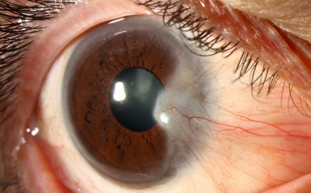
Pterygium grading describes a pterygium by its translucency and by how far its head extends onto the cornea.
Both features help predict recurrence after excision.
They also guide the timing of surgery and the choice of technique.
Why Pterygium Grading Matters in Clinic
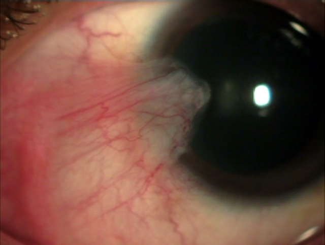
A pterygium is a wing-shaped fibrovascular growth of bulbar conjunctiva onto the cornea.
Patients often call it surfer’s eye because chronic ultraviolet exposure drives it.
However, two lesions of similar size can behave very differently after surgery.
Therefore, a short grade in the chart carries more information than the diagnosis alone.
The Tan Morphologic Classification
Tan and colleagues graded pterygia by how well the episcleral vessels show through the lesion body.
They assessed each eye at the slit lamp before excision.
Then they related each grade to recurrence in a controlled surgical trial.
The system needs no imaging and takes seconds to apply.
All-fit Slit-Lamp Adapter
Record and share exactly what you see at the slit lamp. One adapter fits any slit lamp or surgical microscope — and any smartphone.
From Choroida — the team behind this siteTan Stages in Pterygium Grading
- T1 (atrophic): episcleral vessels are clearly visible under the body
- T2 (intermediate): episcleral vessels are partly visible
- T3 (fleshy): thick tissue hides the episcleral vessels completely
How to Grade Translucency at the Slit Lamp
Use diffuse white light with moderate magnification.
Next, focus on the body of the lesion, away from the head and cap.
Look for the fine radial episcleral vessels beneath the conjunctival tissue.
If findings fall between two grades, note both and recheck at the next visit.
Why Fleshy Lesions Recur
A fleshy pterygium probably reflects more active fibrovascular proliferation.
In addition, thick lesions commonly appear in younger patients with ongoing sun exposure.
As a result, any residual tissue may regrow quickly after excision.
Pterygium Grading and Recurrence Risk
In the Tan trial, fleshy pterygia recurred more often than atrophic ones after bare sclera excision.
Conjunctival autografting reduced recurrence compared with bare sclera.
Therefore, a T3 grade argues firmly against bare sclera surgery (see conjunctival autograft technique).
Corneal Extent in Pterygium Grading
A second system measures how far the head advances toward the pupil.
- Grade I: head between the limbus and a point midway to the pupil margin
- In grade II, the head lies between that midpoint and the pupil margin
- For grade III, the head crosses the pupil margin
Some authors use slightly different cut points, so state the system you follow.
Measuring Extent Accurately
Measure horizontal encroachment from the limbus in millimeters with the slit beam.
Also photograph the eye at each visit for objective comparison.
Corneal topography adds useful data because pterygia distort the corneal surface.
Astigmatism and Visual Impact
As the head advances, it flattens the cornea along the horizontal meridian.
This flattening produces with-the-rule astigmatism that glasses may not fully correct.
Later, the lesion may enter the visual axis and block vision directly.
Pterygium Grading and Surgical Timing
Small atrophic lesions without symptoms usually need observation and ultraviolet protection.
However, consider surgery when the head approaches the pupil margin or astigmatism progresses.
Chronic irritation and cosmetic concern are also valid indications.
Planning Cataract or Refractive Surgery
A significant pterygium distorts keratometry and can shift intraocular lens calculations.
Therefore, remove it first and wait for corneal stability before repeating biometry.
Then plan the lens surgery on stable measurements (see cataract surgery timing).
Choosing the Technique by Grade
Conjunctival autograft is the preferred approach for most primary lesions.
Fibrin glue shortens surgery and improves comfort compared with sutures (see fibrin glue grafts).
For high-risk fleshy lesions, some surgeons add mitomycin C.
However, mitomycin C can cause scleral thinning and melt, so use it cautiously.
Recurrent Lesions
Recurrent pterygia are often fleshier and adhere more firmly to deeper tissue.
They may also restrict ocular motility through scarring.
Grade them with the same scales, and record the number of prior surgeries (see pterygium recurrence).
Differential Diagnosis Before Pterygium Grading
- Pinguecula: yellowish nodule that stays off the cornea
- Pseudopterygium: follows injury or ulceration, and a probe passes beneath it
- Ocular surface squamous neoplasia: gelatinous or leukoplakic lesion with feeder vessels
- Limbal dermoid: congenital, smooth and often with hairs
Pterygium Grading in Patient Counseling
Explain the grade in plain language at the first visit.
For example, tell patients that a fleshy lesion carries a higher chance of coming back.
Also advise sunglasses and wide-brimmed hats to reduce ultraviolet exposure.
Finally, explain that surgery can fail even with the best technique.
Follow-Up and Documentation
Record the Tan grade, corneal extent and astigmatism at each visit.
Next, compare serial photographs to detect real growth.
After surgery, watch for early fleshy regrowth during the first months.
Pitfalls to Avoid
- Do not grade translucency from the head alone
- Never ignore an atypical, gelatinous or leukoplakic lesion
- Avoid bare sclera excision for fleshy pterygia
- Do not perform biometry with a significant pterygium in place


Document what you see
Two smartphone imaging tools built for everyday clinic use — one for the slit lamp, one for the fundus.
From Choroida — the team behind this siteReferences
- Tan DT, Chee SP, Dear KB, Lim AS. Effect of pterygium morphology on pterygium recurrence in a controlled trial comparing conjunctival autografting with bare sclera excision. Arch Ophthalmol. 1997;115:1235-1240.
- Hirst LW. The treatment of pterygium. Surv Ophthalmol. 2003;48:145-180.
- Kaufman SC, Jacobs DS, Lee WB, et al. Options and adjuvants in surgery for pterygium: a report by the American Academy of Ophthalmology. Ophthalmology. 2013;120:201-208.
- Kenyon KR, Wagoner MD, Hettinger ME. Conjunctival autograft transplantation for advanced and recurrent pterygium. Ophthalmology. 1985;92:1461-1470.
- Salmon JF. Kanski’s Clinical Ophthalmology: A Systematic Approach. 9th ed. Elsevier; 2020.

Writes and reviews the clinical articles on Educate. Every post is checked against peer-reviewed sources before it goes live.