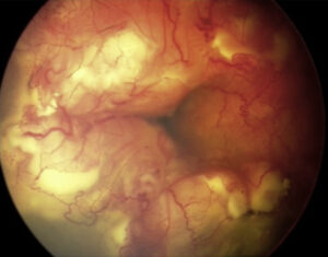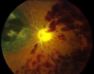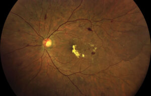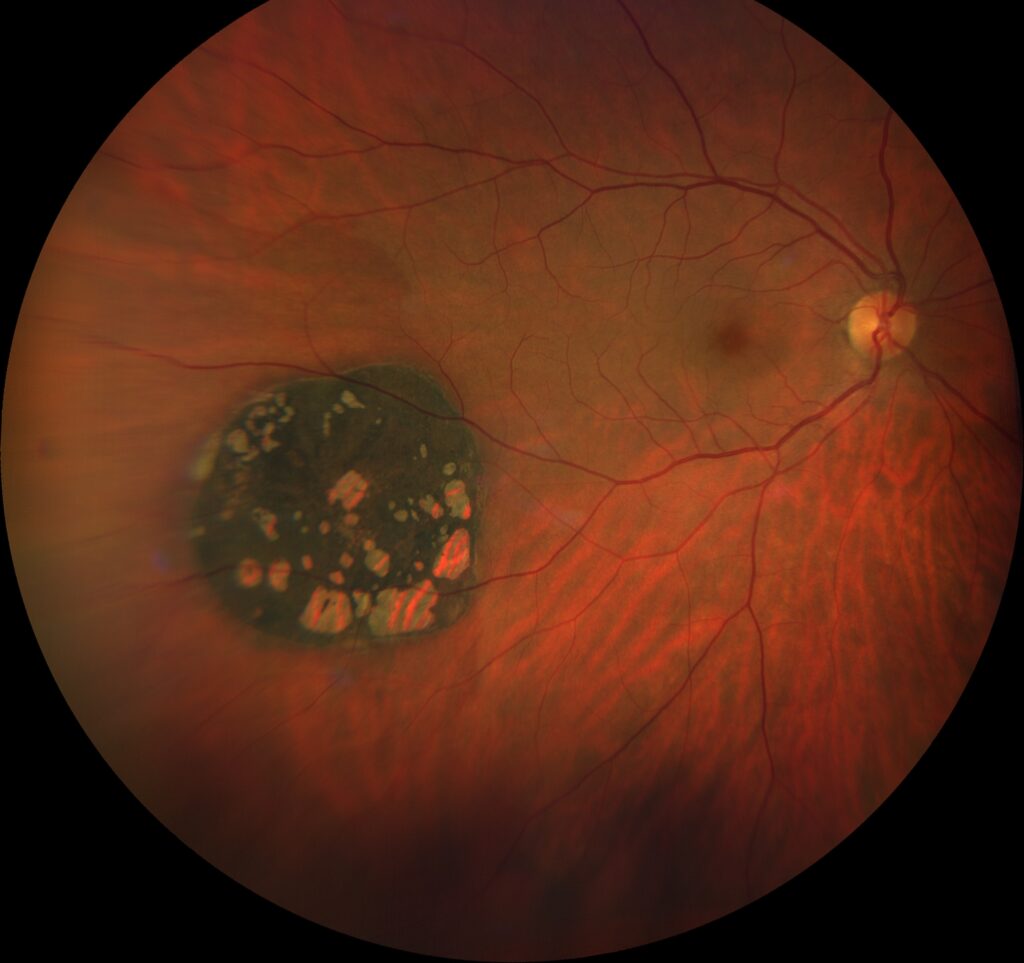
Pigmented fundus lesions are dark spots found on fundus examination, and most of them are benign.
However, a missed small melanoma can cost the eye or the patient’s life, and therefore every dark lesion deserves a structured description and baseline imaging.
Where Pigmented Fundus Lesions Arise
The layer of origin predicts much of the behavior. Nevi and melanomas arise from choroidal melanocytes beneath the retinal pigment epithelium (RPE), while CHRPE and reactive hyperplasia start in the RPE itself. As a result, depth and color on examination already narrow the differential.
Choroidal Nevus
A typical nevus is flat or slightly elevated and slate gray, with feathery but defined borders. Overlying drusen suggest chronicity because the RPE has had time to degenerate, and for transformation risk, see TFSOM risk scoring.
Fundus Explorer Pro
Photograph the retinal findings described here with the phone already in your pocket — 22 D optics and built-in illumination in one handheld unit.
From Choroida — the team behind this siteChoroidal Melanoma
Melanoma tends to be thicker, with a dome shape or a mushroom shape after it breaks through Bruch membrane. Its color ranges from brown to amelanotic, so absent pigment does not exclude it, and orange lipofuscin pigment and subretinal fluid over the surface are warning signs.
Risk Factors in Pigmented Fundus Lesions
Shields and colleagues summarized nevus transformation risk with the mnemonic TFSOM-DIM.
- Thickness over 2 mm on ultrasonography
- Subretinal fluid on OCT
- Symptoms such as reduced vision
- Orange pigment on autofluorescence
- Melanoma acoustic hollowness on ultrasonography
- Diameter over 5 mm on photography
Each added factor raises the estimated risk, so multiple factors justify oncology review.
CHRPE
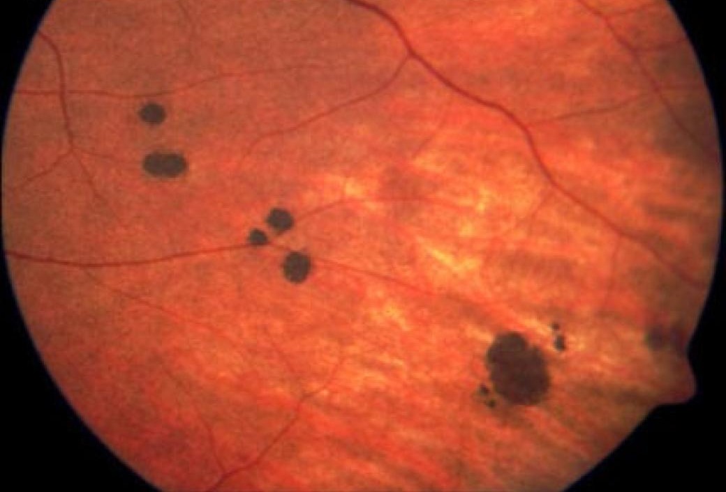
Solitary CHRPE is flat, jet black and sharply demarcated, often in the midperiphery, and depigmented lacunae and a pale margin develop as it ages. Slow enlargement is common and does not, by itself, signal malignancy, but multiple irregular fishtail lesions in both eyes suggest familial adenomatous polyposis (see CHRPE overview).
Melanocytoma of the Optic Disc
Melanocytoma is a jet-black lesion that usually overlies the optic disc, and its edges look fibrillated because the cells extend into the nerve fiber layer. Vision is often good, although a relative afferent pupillary defect may occur. Malignant transformation is rare, so periodic photographs are reasonable (see choroidal melanocytoma).
Reactive RPE Hyperplasia
Hyperplasia appears as irregular black clumps or plaques with uneven borders. It follows trauma, inflammation, laser, retinal detachment or chronic subretinal fluid, so look for the cause, such as chorioretinal scars or demarcation lines.
Comparing Pigmented Fundus Lesions
- Choroidal nevus: slate gray, flat to under 2 mm, drusen and halo common
- Choroidal melanoma: brown or amelanotic, thicker, orange pigment and fluid
- Solitary CHRPE: jet black, flat, sharp margin, lacunae
- Melanocytoma: jet black, on the disc, feathery edges
- RPE hyperplasia: black, irregular, with scarring or prior disease
- Subretinal hemorrhage: dark red to black, changes color over weeks
Imaging Pigmented Fundus Lesions
Enhanced depth imaging OCT shows whether the lesion lies in the choroid or the RPE, and it also detects shallow subretinal fluid that the ophthalmoscope misses. Fundus autofluorescence makes orange pigment appear bright, while B-scan measures thickness and A-scan shows the low to medium internal reflectivity typical of melanoma.
Mimics of Pigmented Fundus Lesions
Subretinal or sub-RPE hemorrhage can look black and elevated, as in peripheral exudative hemorrhagic chorioretinopathy, which often mimics melanoma in older adults. In addition, metastasis from cutaneous melanoma may be pigmented, but repeat examination usually separates blood from tumor, because hemorrhage evolves.
When to Refer
Refer promptly for growth, subretinal fluid, orange pigment or thickness over 2 mm. A new pigmented lesion in an adult deserves specialist review too.
Follow-Up of Pigmented Fundus Lesions
For a low-risk nevus, many clinicians repeat photography and OCT within several months. If stable, yearly review is common. However, documented growth turns any lesion into a suspicious one.
Treatment Overview
Benign nevi, CHRPE and hyperplasia need observation only, while for melanoma, options include plaque brachytherapy, proton beam radiotherapy and enucleation, depending on size and location. The COMS found similar long-term mortality after iodine-125 brachytherapy and enucleation for medium tumors. However, local control cannot guarantee freedom from metastasis, so patients also need systemic surveillance, usually including liver imaging.
Counseling Patients About Pigmented Fundus Lesions
Explain that most dark spots behave like benign freckles of the eye, and that change over time drives decisions, so baseline images matter. Finally, ask patients to report new flashes, floaters or blur.
Pitfalls to Avoid
- Do not label a nevus without measuring thickness
- Never ignore shallow fluid seen only on OCT
- Do not assume absent pigment excludes melanoma
- Avoid judging growth from memory instead of photographs


Document what you see
Two smartphone imaging tools built for everyday clinic use — one for the slit lamp, one for the fundus.
From Choroida — the team behind this siteReferences
- Shields CL, Dalvin LA, Ancona-Lezama D, et al. Choroidal nevus imaging features in 3,806 cases and risk factors for transformation into melanoma in 2,355 cases: the 2020 Taylor R. Smith and Victor T. Curtin Lecture. Retina. 2019;39:1840-1851.
- Shields CL, Mashayekhi A, Ho T, et al. Solitary congenital hypertrophy of the retinal pigment epithelium: clinical features and frequency of enlargement in 330 patients. Ophthalmology. 2003;110:1968-1976.
- Shields JA, Demirci H, Mashayekhi A, Shields CL. Melanocytoma of optic disc in 115 cases: the 2004 Samuel Johnson Memorial Lecture, part 1. Ophthalmology. 2004;111:1739-1746.
- Collaborative Ocular Melanoma Study Group. The COMS randomized trial of iodine 125 brachytherapy for choroidal melanoma: V. Twelve-year mortality rates and prognostic factors: COMS report No. 28. Arch Ophthalmol. 2006;124:1684-1693.
Test yourself
A few questions straight from this article.
-
From which cells do choroidal nevi and choroidal melanomas arise?
Nevi and melanomas arise from choroidal melanocytes beneath the RPE, whereas CHRPE and reactive hyperplasia start in the RPE itself, so depth already narrows the differential. -
What do drusen overlying a choroidal nevus suggest?
Overlying drusen suggest chronicity because the RPE over the nevus has had time to degenerate. -
In the TFSOM-DIM risk mnemonic for choroidal nevus transformation, what ultrasound thickness counts as a risk factor?
Thickness over 2 mm on ultrasonography is a TFSOM-DIM factor and, on its own, a reason for prompt referral. -
In the TFSOM-DIM mnemonic, what photographic diameter of a choroidal nevus counts as a risk factor?
Diameter over 5 mm on photography is one TFSOM-DIM factor; each added factor raises the estimated risk of transformation. -
Which imaging modality makes orange lipofuscin pigment over a choroidal lesion appear bright?
Fundus autofluorescence shows orange pigment as bright, while B-scan measures thickness and EDI-OCT locates the lesion and detects shallow fluid. -
What shape does a choroidal melanoma typically take after it breaks through Bruch membrane?
Melanoma tends to be thicker and dome-shaped, becoming mushroom-shaped once it breaks through Bruch membrane. -
Multiple irregular fishtail-shaped CHRPE-like lesions in both eyes should raise suspicion of which systemic condition?
Solitary CHRPE is benign, but multiple irregular fishtail lesions in both eyes suggest familial adenomatous polyposis. -
Which jet-black pigmented lesion usually overlies the optic disc and may cause a relative afferent pupillary defect?
Melanocytoma usually overlies the disc with edges extending into the nerve fibre layer; vision is often good and malignant transformation is rare. -
How is a dark subretinal hemorrhage usually separated from a pigmented choroidal tumor?
Hemorrhage, as in peripheral exudative hemorrhagic chorioretinopathy, changes colour over weeks, so repeat examination usually separates blood from tumor. -
For medium choroidal melanomas, how did long-term mortality compare after iodine-125 brachytherapy versus enucleation in the COMS?
The COMS found similar long-term mortality after iodine-125 brachytherapy and enucleation; local control does not prevent metastasis, so systemic surveillance is still needed.

Writes and reviews the clinical articles on Educate. Every post is checked against peer-reviewed sources before it goes live.