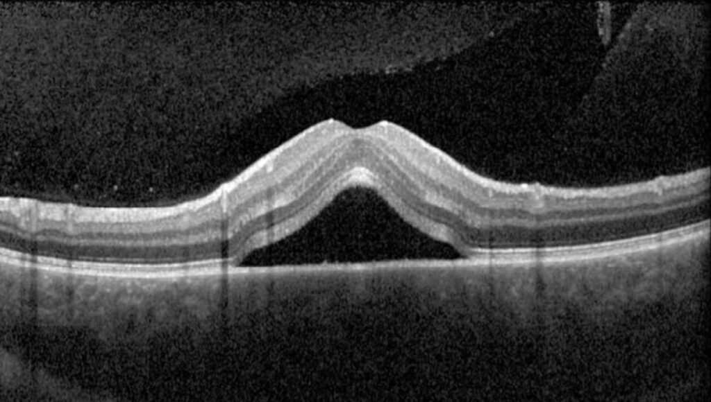
Intraretinal fluid appears on OCT as dark, hyporeflective spaces within the neurosensory retina.
Clinicians call these spaces cysts, although they lack an epithelial lining.
However, some dark spaces reflect tissue loss and contain no active leakage.
Therefore, decide whether the space is exudative or degenerative before changing treatment.
What Intraretinal Fluid Looks Like on OCT
Exudative fluid usually forms round or oval spaces with fairly sharp borders.
These spaces commonly sit in the inner nuclear and outer plexiform layers.
Also, dense raster scans can reveal small cysts that a single foveal line misses.
Why Fluid Collects in the Retina
Fluid accumulates when entry into the retina exceeds removal.
For example, a damaged blood-retinal barrier increases leakage from vessels.
Meanwhile, Müller cells and the retinal pigment epithelium help remove fluid, so their dysfunction may worsen edema.
Fundus Explorer Pro
Photograph the retinal findings described here with the phone already in your pocket — 22 D optics and built-in illumination in one handheld unit.
From Choroida — the team behind this siteSymptoms and Examination Clues
Patients typically report blur, reduced contrast or distorted lines.
Because subtle edema often escapes biomicroscopy, OCT usually confirms the finding.
Intraretinal Fluid Versus Subretinal Fluid
Intraretinal spaces lie within the retinal layers, while subretinal fluid lifts the retina from the pigment epithelium.
Both can coexist, especially in neovascular disease and vein occlusion.
Therefore, record each compartment separately at every visit.
Signs of Exudative Intraretinal Fluid
First, the retina is usually thickened, and the cysts change between visits.
Next, look for hard exudates, hemorrhage or hyperreflective foci nearby.
Fluorescein leakage or a visible neovascular lesion then adds support.
Signs of Degenerative Spaces
Degenerative cavities often have angular or irregular outlines.
In addition, the surrounding retina tends to be thin.
They frequently overlie atrophy or marked ellipsoid zone disruption.
Because tissue loss creates these spaces, they often resist anti-VEGF therapy.
Outer Retinal Tubulation and Intraretinal Fluid
Outer retinal tubulation is a common mimic in advanced AMD.
On B-scan, it forms round or ovoid spaces with hyperreflective borders.
Therefore, repeated injections for this finding alone would expose the patient to needless treatment.
Macular Telangiectasia and Other Mimics
Macular telangiectasia type 2 can produce foveal cavities without the expected retinal thickening.
Gaudric and colleagues documented this mismatch.
Also, X-linked retinoschisis and myopic foveoschisis cause splitting that can resemble cystoid edema.
Vitreomacular traction may also create cysts, so inspect the vitreoretinal interface.
Confirming Activity With Angiography
Fluorescein angiography shows leakage and, in cystoid edema, late petaloid pooling.
However, it cannot separate retinal layers as OCT does.
OCT angiography can reveal a neovascular network without dye, although it does not show leakage.
Then compare current scans with earlier ones.
Intraretinal Fluid in Neovascular AMD
In neovascular AMD, foveal cysts generally carry an unfavorable visual association.
For example, the CATT morphology analysis linked intraretinal fluid with worse acuity.
Waldstein and colleagues also reported a stronger link between acuity and intraretinal volume than subretinal volume.
Nevertheless, these associations cannot predict one patient’s outcome.
Diabetic, Vein Occlusion and Inflammatory Edema
Diabetic macular edema and retinal vein occlusion commonly cause cystoid spaces.
Because ischemia and photoreceptor damage also limit recovery, a dry macula may still see poorly.
Cystoid edema after cataract surgery or uveitis can look similar on OCT.
In these settings, inflammation guides therapy, and anti-VEGF may be unnecessary.
Treating Intraretinal Fluid
Anti-VEGF therapy reduces vascular leakage and treats the driving lesion in neovascular AMD.
However, it does not restore photoreceptors that have already been lost.
The FLUID trial tolerated some subretinal fluid while still retreating all intraretinal fluid.
Its 24-month visual outcomes were similar between groups, although the result applies to that protocol.
Steroids and Other Options
Corticosteroids may help selected uveitic, diabetic or vein occlusion edema.
However, they can raise intraocular pressure and accelerate cataract.
If traction is the cause, then vitrectomy may be considered in selected eyes.
Counseling and Follow-Up
Explain that the scan shows fluid or tissue change, which vision may not mirror exactly.
Also, tell patients with degenerative cavities why more injections would not help.
At follow-up, compare identical scan locations, thickness, acuity and outer retinal integrity.
Pitfalls to Avoid
- Do not treat every dark space as active leakage
- Never rely on one foveal B-scan alone
- Do not overlook traction, drug toxicity or inflammation
- Compare old scans before you escalate therapy


Document what you see
Two smartphone imaging tools built for everyday clinic use — one for the slit lamp, one for the fundus.
From Choroida — the team behind this siteReferences
- Spaide RF. Retinal vascular cystoid macular edema: review and new theory. Retina. 2016;36:1823-1842.
- Jaffe GJ, Martin DF, Toth CA, et al. Macular morphology and visual acuity in the Comparison of Age-related Macular Degeneration Treatments Trials. Ophthalmology. 2013;120:1860-1870.
- Waldstein SM, Philip AM, Leitner R, et al. Correlation of 3-dimensionally quantified intraretinal and subretinal fluid with visual acuity in neovascular age-related macular degeneration. JAMA Ophthalmol. 2016;134:182-190.
- Zweifel SA, Engelbert M, Laud K, et al. Outer retinal tubulation: a novel optical coherence tomography finding. Arch Ophthalmol. 2009;127:1596-1602.
- Gaudric A, Ducos de Lahitte G, Cohen SY, et al. Optical coherence tomography in group 2A idiopathic juxtafoveolar retinal telangiectasis. Arch Ophthalmol. 2006;124:1410-1419.
- Guymer RH, Markey CM, McAllister IL, et al. Tolerating subretinal fluid in neovascular age-related macular degeneration treated with ranibizumab using a treat-and-extend regimen: FLUID study 24-month results. Ophthalmology. 2019;126:723-734.

Writes and reviews the clinical articles on Educate. Every post is checked against peer-reviewed sources before it goes live.