Ring scotoma is a circular band of field loss surrounding fixation, with vision preserved inside it.
Patients often experience it as early tunnel vision, although many first notice bumping into objects.
Retinitis pigmentosa is the classic cause, while retinal toxicity, glaucoma and optics can mimic it.
What a Ring Scotoma Looks Like
The defect usually lies in the midperiphery, roughly 20 to 50 degrees from fixation.
Early loss may appear as separate patches, which then join into a partial or complete circle.
Central acuity can stay normal, so the letter chart may falsely reassure both patient and examiner.
Why Rod Loss Comes First
Rod density peaks about 20 degrees from the fovea, within the zone that often fails first.
In rod-cone dystrophies, rods degenerate early, and then cones decline secondarily.
As a result, midperipheral function falls while the cone-rich macula keeps working into the later stages of disease.
However, topography alone cannot explain every field, because genotype and disease stage also shape the pattern.
Fundus Explorer Pro
Photograph the retinal findings described here with the phone already in your pocket — 22 D optics and built-in illumination in one handheld unit.
From Choroida — the team behind this siteRing Scotoma in Retinitis Pigmentosa
Patients typically report night blindness first, and then gradual loss of side vision.
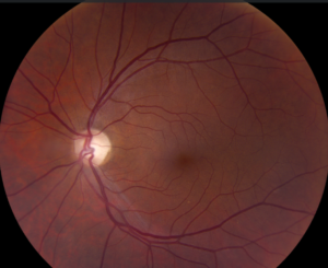
The fundus may show bone-spicule pigment, arteriolar attenuation and waxy disc pallor.
Over time, the ring expands outward to the periphery and also inward toward fixation.
Finally, a small central tunnel may remain, sometimes with a separate peripheral island.
Other Retinal Causes of Ring Scotoma
When the fundus lacks classic signs, widen the differential (see other causes of night vision loss).
- Hydroxychloroquine retinopathy, especially with a pericentral pattern
- Autoimmune retinopathy, including cancer-associated retinopathy, with faster progression
- Choroideremia, with progressive chorioretinal atrophy in affected males
- Gyrate atrophy, with scalloped peripheral atrophy and raised ornithine
- Vitamin A deficiency, which can improve after replacement
Hydroxychloroquine and the Pericentral Ring
Melles and Marmor described pericentral toxicity, which appeared more often in Asian patients.
Therefore, a 10-2 field alone can miss early damage in these patients.
AAO screening recommendations advise wider testing, such as 24-2 or 30-2 fields, for Asian patients.
Glaucoma Can Mimic a Ring Scotoma
Advanced glaucoma can produce superior and inferior arcuate defects that nearly encircle fixation.
However, these defects follow nerve fiber bundles and often respect the horizontal nasal meridian.
Next, compare the field with disc cupping and retinal nerve fiber layer thinning.
Optical Ring Scotoma From High Plus Lenses
Strong plus spectacles, classically for aphakia, create a ring-shaped blind zone near the lens edge.
This scotoma moves with eye movement, and objects may suddenly pop into view.
Because the retina is healthy, a contact lens or intraocular lens removes the problem.
Night Tasks and Dark Adaptation
Rod dysfunction slows dark adaptation and reduces sensitivity in dim settings.
For example, patients struggle when entering a dark cinema or walking outdoors at dusk.
Night driving often becomes difficult early, sometimes before the patient recognizes any field loss.
Also, ask directly about these tasks, because patients may blame their glasses or aging.
Mobility Problems With Ring Scotoma
Midperipheral loss hides obstacles that normally attract attention from the side.
As a result, patients may miss curbs, low furniture, stair edges and approaching people.
Acuity often predicts these difficulties poorly, while field extent usually gives more useful information.
Geruschat and colleagues also examined mobility performance directly in patients with retinitis pigmentosa.
Testing a Ring Scotoma
Goldmann kinetic perimetry maps the whole ring, especially with several isopters such as V4e and III4e.
In contrast, a 10-2 or 24-2 program may show only fragments of the defect.
Then repeat testing with the same method, because strategy changes can imitate progression.
Imaging and Electrophysiology
OCT often shows ellipsoid zone loss in the midperiphery, with a preserved central island.
Autofluorescence may reveal a hyperautofluorescent ring around the macula in retinitis pigmentosa.
Full-field ERG can then confirm generalized rod and cone dysfunction, while multifocal ERG helps assess pericentral and central disease.
Ring Scotoma Treatment by Cause
Treatment depends entirely on the cause, so a diagnosis should come first.
Stopping hydroxychloroquine, in coordination with the prescriber, limits further exposure, although damage may still progress.
Voretigene neparvovec improved multi-luminance mobility testing in biallelic RPE65 disease, yet it suits only patients with viable retinal cells.
Also, treat cataract and cystoid macular edema in retinitis pigmentosa, because both can reduce remaining vision.
Counseling Patients With Ring Scotoma
Explain that normal acuity does not exclude serious field loss.
In addition, discuss driving rules, lighting, orientation and mobility training, and genetic counseling where relevant.
Refer early for rehabilitation, because some low vision strategies also help inherited retinal disease.
At follow-up, review fields, macular OCT and changes in daily function.
Pitfalls to Avoid
- Do not dismiss night complaints because acuity measures 20/20
- Never rely on a central field alone when symptoms suggest peripheral loss
- Do not miss rapid progression, which should prompt autoimmune evaluation
- Do not label every ring as retinitis pigmentosa without ERG or genetic testing


Document what you see
Two smartphone imaging tools built for everyday clinic use — one for the slit lamp, one for the fundus.
From Choroida — the team behind this siteReferences
- Hartong DT, Berson EL, Dryja TP. Retinitis pigmentosa. Lancet. 2006;368:1795-1809.
- Melles RB, Marmor MF. Pericentral retinopathy and racial differences in hydroxychloroquine toxicity. Ophthalmology. 2015;122:110-116.
- Marmor MF, Kellner U, Lai TY, et al. Recommendations on screening for chloroquine and hydroxychloroquine retinopathy (2016 revision). Ophthalmology. 2016;123:1386-1394.
- Russell S, Bennett J, Wellman JA, et al. Efficacy and safety of voretigene neparvovec (AAV2-hRPE65v2) in patients with RPE65-mediated inherited retinal dystrophy. Lancet. 2017;390:849-860.
- Geruschat DR, Turano KA, Stahl JW. Traditional measures of mobility performance and retinitis pigmentosa. Optom Vis Sci. 1998;75:525-537.

Writes and reviews the clinical articles on Educate. Every post is checked against peer-reviewed sources before it goes live.