Calcified drusen are mineralized sub-RPE deposits that appear as glistening, refractile spots in age-related macular degeneration (AMD).
They usually occur in eyes with established intermediate disease.
However, they carry a recognized association with later atrophy.
What Are Calcified Drusen?
Drusen accumulate between the RPE and Bruch membrane, and some mineralize over time.
Histologic studies have identified hydroxyapatite spherules within these deposits.
However, a clinical label only infers composition, because we cannot biopsy a druse in clinic.
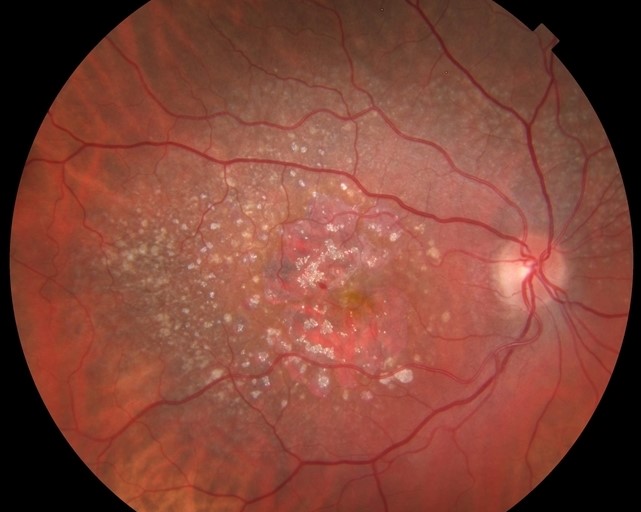
Fundus Appearance of Calcified Drusen
At the slit lamp, look for small bright points with a crystalline, sparkling quality.
These points usually sit within a yellow druse (see soft and hard drusen).
Nearby pigment clumping is also common.
Therefore, examine the whole macula at high magnification.
Fundus Explorer Pro
Photograph the retinal findings described here with the phone already in your pocket — 22 D optics and built-in illumination in one handheld unit.
From Choroida — the team behind this siteWhy the Spots Glisten
Mineral probably reflects and scatters light differently from the surrounding lipid-rich material.
As a result, the examiner sees a sharp glint against a duller yellow background.
The exact optical explanation remains uncertain.
Symptoms and History
Calcified deposits produce no specific symptom.
Instead, patients report the complaints of underlying AMD, such as difficulty reading in dim light.
However, new distortion or sudden central blur should prompt a search for neovascularization.
Imaging Calcified Drusen
Color photography documents the refractile quality and allows comparison between visits.
Fundus autofluorescence is more variable, because it reflects RPE health more than the deposit itself.
Therefore, autofluorescence supports the assessment but cannot identify calcification alone.
OCT Features of Calcified Drusen
Structural OCT typically shows a druse with a hyporeflective interior and a hyperreflective border.
The overlying RPE and outer retina often look thinned or disrupted.
In addition, focal hypertransmission may appear beneath the lesion.
Next, correlate each finding with the color photograph, since no single B-scan sign proves mineralization.
En Face OCT for Calcified Drusen
Swept-source en face imaging can map these lesions across the macula.
A slab beneath the RPE reveals bright hypertransmission foci.
Then, B-scans through each focus help separate calcified drusen from early atrophy.
Availability limits routine use.
Hyporeflective Cores and Progression
Hyporeflective core drusen have been linked with progression to geographic atrophy.
Calcific material may account for some of these low-reflectivity interiors.
However, other contents can look similar, so a dark core does not establish calcification.
Calcified Drusen and Atrophy Risk
Refractile deposits have been associated with later macular atrophy in observational AMD cohorts.
These studies show association, so they do not prove that mineral causes RPE death.
Also, published data do not support a validated individual risk score.
Why Atrophy May Follow
Calcification may mark a late stage in the drusen life cycle.
Overlying RPE cells can dysfunction or die as the deposit matures.
Photoreceptors then lose support (see ellipsoid zone disruption).
Differential Diagnosis of Calcified Drusen
Several conditions also produce bright or crystalline deposits.
- Hard exudates, which lie within the retina and accompany vascular leakage
- Macular telangiectasia type 2, with superficial crystals and OCT cavities
- Bietti crystalline dystrophy, with widespread crystals and chorioretinal atrophy
- Tamoxifen or canthaxanthin retinopathy, identified by medication history
- Talc retinopathy, with perivascular refractile emboli
What Changes the Diagnosis
Young age should prompt consideration of inherited drusen disease.
Similarly, inner retinal crystals point away from a sub-RPE process.
Finally, hemorrhage or subretinal fluid requires angiography to exclude neovascularization.
Managing Calcified Drusen
No treatment removes the mineral or restores overlying tissue.
Therefore, management follows the AMD stage.
AREDS2 supplements suit eligible patients with intermediate disease, although they do not target calcification.
Complement inhibitors apply only once geographic atrophy develops (see geographic atrophy treatment).
They slow lesion growth, but they do not restore lost vision.
Follow-Up and Monitoring
Serial OCT should look for hypertransmission, RPE loss and outer retinal collapse.
The CAM consensus criteria give a shared language for describing atrophy.
In addition, check for neovascular changes, because these eyes still have AMD.
Counseling the Patient
First, explain that these spots are aging deposits unrelated to dietary calcium.
Next, describe them as a possible marker of higher atrophy risk, without promising a timeline.
Smoking cessation is the most useful modifiable step.
Finally, teach the patient which new symptoms need a prompt visit.
Pitfalls to Avoid
- Do not call every glistening macular spot a calcified druse
- Avoid diagnosing calcification from a hyporeflective core alone
- Do not confuse hypertransmission with complete RPE atrophy
- Never skip the medication history when crystals look atypical


Document what you see
Two smartphone imaging tools built for everyday clinic use — one for the slit lamp, one for the fundus.
From Choroida — the team behind this siteReferences
- Tan ACS, Pilgrim MG, Fearn S, et al. Calcified nodules in retinal drusen are associated with disease progression in age-related macular degeneration. Sci Transl Med. 2018;10:eaat4544.
- Suzuki M, Curcio CA, Mullins RF, Spaide RF. Refractile drusen: clinical imaging and candidate histology. Retina. 2015;35:859-865.
- Oishi A, Thiele S, Nadal J, et al. Prevalence, natural course, and prognostic role of refractile drusen in age-related macular degeneration. Invest Ophthalmol Vis Sci. 2017;58:2198-2206.
- Veerappan M, El-Hage-Sleiman AM, Tai V, et al. Optical coherence tomography reflective drusen substructures predict progression to geographic atrophy in age-related macular degeneration. Ophthalmology. 2016;123:2554-2570.
- Sadda SR, Guymer R, Holz FG, et al. Consensus definition for atrophy associated with age-related macular degeneration on OCT: Classification of Atrophy Report 3. Ophthalmology. 2018;125:537-548.

Writes and reviews the clinical articles on Educate. Every post is checked against peer-reviewed sources before it goes live.