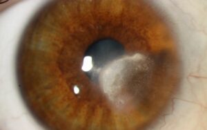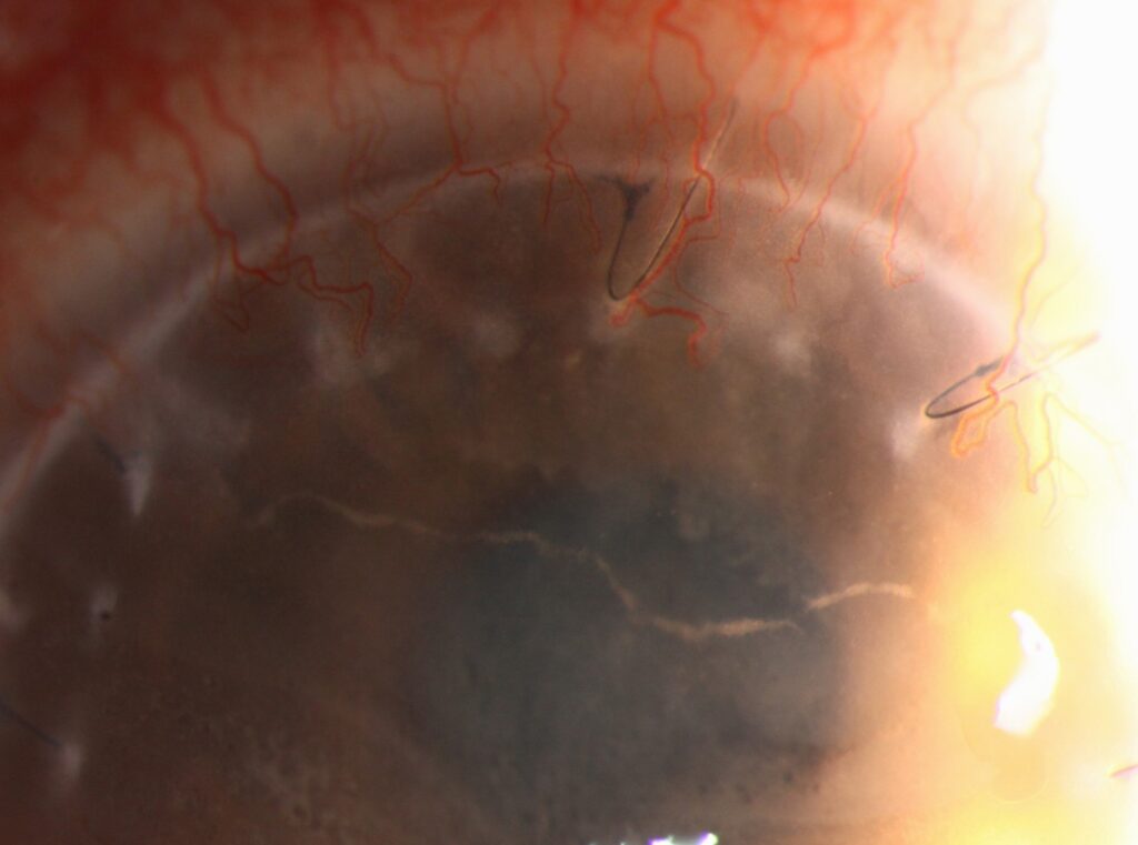
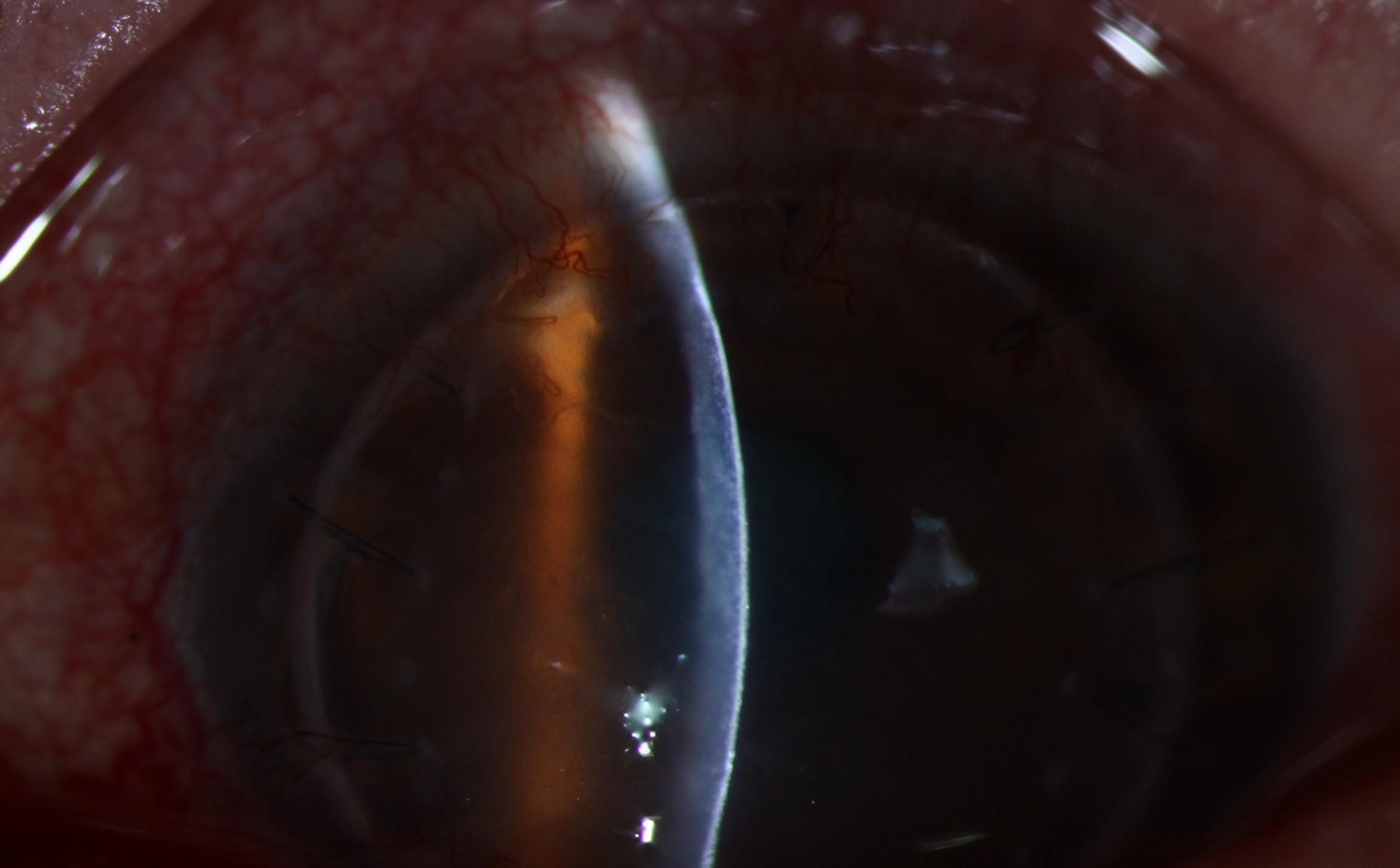
Khodadoust line describes a band of keratic precipitates that crosses the endothelium of a corneal graft during acute rejection.
It usually follows penetrating keratoplasty.
Therefore, this sign calls for same-day assessment and intensive steroid treatment.
What the Khodadoust Line Is
The line consists of lymphocytes and other inflammatory cells adherent to the donor endothelium.
These cells damage donor endothelial cells as the front advances.
The name honors Ali Khodadoust, whose work with Silverstein defined rejection of individual corneal layers.
Slit Lamp Appearance of a Khodadoust Line
Look for a thin white or gray line of fine precipitates on the posterior graft surface.
It often looks curved, with a distinct advancing edge.
The stroma behind the line looks edematous, while the cornea ahead may remain clear.
In addition, the anterior chamber often shows mild cells and flare.
All-fit Slit-Lamp Adapter
Record and share exactly what you see at the slit lamp. One adapter fits any slit lamp or surgical microscope — and any smartphone.
From Choroida — the team behind this siteHow a Khodadoust Line Progresses
The front usually starts at the graft-host junction, often near vascularized host tissue.
Without treatment, the line typically moves across the graft over days.
Its speed varies between patients, so a single examination cannot predict the pace.
Human endothelial cells regenerate poorly, so cell loss is largely permanent.
As a result, repeated episodes raise the risk of corneal graft failure.
Symptoms and Risk Factors
Patients describe blurred vision, redness, light sensitivity or a dull ache.
However, early symptoms can be mild, and vision may seem nearly normal.
For that reason, many surgeons teach graft patients the RSVP warning signs: redness, sensitivity, vision loss and pain.
Stromal vascularization, previous rejection and loose sutures all raise the risk (see early warning signs).
Also, ask about recent steroid withdrawal and poor drop adherence.
Examining a Suspected Khodadoust Line
First, use a narrow slit beam to confirm the endothelial level of the deposits.
Next, use retroillumination to trace the line.
Then measure intraocular pressure and grade the anterior chamber reaction.
Pachymetry or anterior segment OCT can document thickness for comparison.
Finally, a serial photograph helps show whether the front has stopped moving.
Differential Diagnosis of a Khodadoust Line
Herpetic endotheliitis can closely mimic this sign.
Cytomegalovirus deserves suspicion when rejection recurs or fails to respond to steroids.
In addition, uveitis can scatter precipitates, although they usually lack an advancing front.
An epithelial rejection line differs because it lies on the surface and stains with fluorescein.
Other patterns appear in our review of the four types.
Rejection After DSAEK and DMEK
The classic line is mainly associated with penetrating keratoplasty.
After endothelial keratoplasty, rejection more often presents with scattered precipitates and subtle edema.
Therefore, the absence of a line does not exclude rejection in these eyes.
Reported rejection rates also appear lower after DMEK than after DSAEK or penetrating keratoplasty.
Treating a Khodadoust Line
Treatment should begin on the day of diagnosis.
Delayed treatment allows the front to cover more endothelium, and reversal becomes less likely.
Intensive topical steroid forms the base, commonly prednisolone acetate 1% hourly while awake.
The drug suppresses the immune attack, but it cannot replace cells already destroyed.
A cycloplegic may also relieve pain.
Start therapy first, then arrange urgent specialist review when the diagnosis looks clear.
Once the line stops and edema improves, taper the steroid slowly.
Systemic Options
Severe or recurrent episodes may justify oral prednisone or intravenous methylprednisolone.
Hill and colleagues compared oral therapy with a single intravenous pulse and reported favorable pulse results.
However, the optimal regimen remains uncertain.
Screen for diabetes, infection and other systemic contraindications before prescribing these drugs.
Risks of Steroid Treatment
Steroids can raise intraocular pressure, worsen herpetic disease and accelerate cataract.
Therefore, check pressure at each visit.
Successful treatment can clear the line and the edema, yet endothelial cell loss may persist.
Persistent edema after adequate treatment suggests irreversible decompensation and may lead to regrafting.
Counseling Patients With a Khodadoust Line
Explain that the immune system is attacking the donor layer that keeps the cornea clear.
Tell patients that many episodes respond when treatment starts early, although recovery varies.
Also, stress frequent drops and warn against stopping steroids suddenly.
Then ask them to return immediately if redness, pain or blur worsens.
Follow-Up After the Line Clears
Review the patient within days until the front stops.
Afterward, extend intervals gradually while tapering therapy.
Each visit should include pressure, pachymetry and a search for new precipitates.
Specular microscopy can later estimate remaining cell density when the view allows.
Pitfalls to Avoid
- Do not dismiss a fine line because vision remains good
- Never taper steroids before the front has stopped
- Do not overlook viral endotheliitis in steroid-resistant cases
- Avoid relying on this line to diagnose rejection after DMEK


Document what you see
Two smartphone imaging tools built for everyday clinic use — one for the slit lamp, one for the fundus.
From Choroida — the team behind this siteReferences
- Khodadoust AA, Silverstein AM. Transplantation and rejection of individual cell layers of the cornea. Invest Ophthalmol. 1969;8:180-195.
- Alldredge OC, Krachmer JH. Clinical types of corneal transplant rejection. Arch Ophthalmol. 1981;99:599-604.
- Panda A, Vanathi M, Kumar A, Dash Y, Priya S. Corneal graft rejection. Surv Ophthalmol. 2007;52:375-396.
- Hill JC, Maske R, Watson P. Corticosteroids in corneal graft rejection: oral versus single pulse therapy. Ophthalmology. 1991;98:329-333.
- Anshu A, Price MO, Price FW. Risk of corneal transplant rejection significantly reduced with Descemet’s membrane endothelial keratoplasty. Ophthalmology. 2012;119:536-540.

Writes and reviews the clinical articles on Educate. Every post is checked against peer-reviewed sources before it goes live.