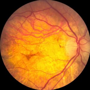
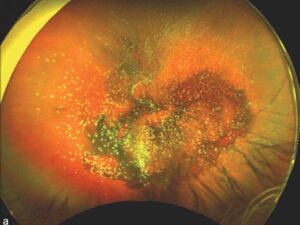
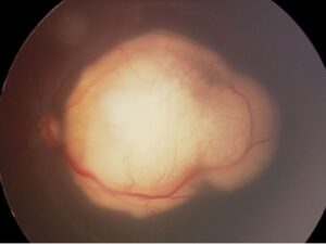
Mizuo phenomenon is a golden or gray-white metallic fundus sheen.
It fades after prolonged dark adaptation and returns after light exposure.
It is the classic sign of Oguchi disease, a form of congenital stationary night blindness.
However, the sheen alone does not establish that diagnosis.
Therefore, examiners should test its behavior in darkness and confirm retinal function with electroretinography.
What Mizuo Phenomenon Looks Like
In the light-adapted state, the fundus shows a shiny yellow to gray sheen with a metallic quality.
Its extent varies, but the reflex often involves the posterior pole and midperiphery.
Meanwhile, the optic disc and vessel caliber usually look normal.
Dark Adaptation in Mizuo Phenomenon
After prolonged darkness, the sheen fades and the fundus regains a more normal color.
Then, light exposure brings the sheen back, often within minutes.
Because routine examination shows only the light-adapted state, the sign needs deliberate retesting.
Fundus Explorer Pro
Photograph the retinal findings described here with the phone already in your pocket — 22 D optics and built-in illumination in one handheld unit.
From Choroida — the team behind this siteWhy the Sheen Appears
The exact mechanism remains uncertain.
Many authors localize the abnormal reflex to the outer retina, where light-dependent changes may alter light scatter.
De Jong and colleagues also described the sign in X-linked retinoschisis.
As a result, any single explanation should be treated as a working hypothesis.
Mizuo Phenomenon and Rod Recovery
Oguchi disease results from biallelic variants in SAG or GRK1.
These genes encode arrestin and rhodopsin kinase, which help shut off activated rhodopsin.
As a result, rods recover very slowly after light exposure.
The disease overview covers inheritance and clinical course (see Oguchi disease).
What the Patient Reports
Most patients describe stable night vision difficulty since early childhood.
Daytime acuity, color vision and peripheral vision are often normal or near normal.
Next, ask about family history and consanguinity, because inheritance is autosomal recessive.
Progressive field loss, however, should prompt a search for another diagnosis.
Confirming Mizuo Phenomenon in Clinic
A simple protocol can document the change without special equipment.
- First, photograph the fundus in the light-adapted state.
- Next, keep the patient in complete darkness for several hours.
- Then, examine or photograph the fundus promptly, using minimal light.
- Finally, reexpose the eye and watch for the sheen to return.
ERG Findings With Mizuo Phenomenon
Full-field ERG supplies the functional evidence.
After standard dark adaptation, the rod response is typically severely reduced or absent.
Also, the bright-flash response often shows an electronegative waveform, while cone responses remain largely preserved.
Prolonged dark adaptation can improve the rod response.
Imaging in Mizuo Phenomenon
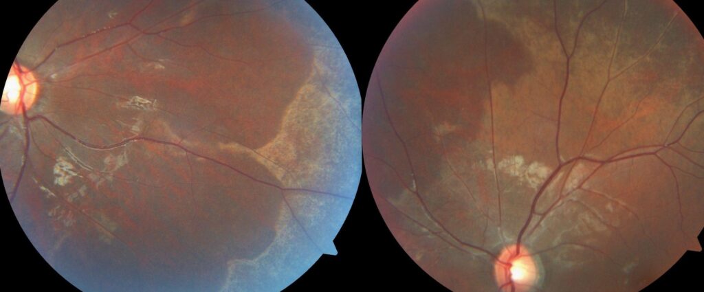
Color photographs provide the simplest record of the sheen before and after darkness.
Meanwhile, OCT may show outer retinal reflectivity changes that vary with adaptation state.
However, no imaging pattern replaces ERG or genetic confirmation.
Differential Diagnosis of a Fundus Sheen
Several conditions can produce a shiny or metallic reflex.
- Normal young retina: a bright internal limiting membrane reflex that moves with the light
- X-linked retinoschisis: foveal schisis, peripheral changes and an electronegative ERG
- Cone dystrophy with tapetal-like sheen: poor central vision and abnormal cone responses
- Fundus albipunctatus: delayed dark adaptation with white dots, without the typical sheen
Comparison With Fundus Albipunctatus
Both conditions can cause lifelong night blindness and improve with prolonged dark adaptation.
However, fundus albipunctatus shows many small white dots and usually lacks the golden sheen (see fundus albipunctatus).
Its mechanism also differs, because it involves the visual cycle.
When Mizuo Phenomenon Is Not Oguchi Disease
The sign is not exclusive to Oguchi disease.
For example, reports describe it in X-linked retinoschisis, where vision loss and foveal changes point elsewhere.
Therefore, reduced acuity, an abnormal cone ERG or progressive field loss should widen the workup.
Managing Patients With Mizuo Phenomenon
The sheen itself needs no treatment, and no therapy has proven benefit for Oguchi disease.
Management therefore centers on accurate diagnosis, genetic counseling and practical advice about night driving and dim environments.
Tell patients that the color change does not itself indicate worsening disease.
Also, explain that prolonged darkness may temporarily improve rod function, although this offers no practical treatment.
Follow-Up and Counseling
Oguchi disease is generally stationary, so many patients need only periodic review.
However, some reports link SAG or GRK1 variants with progressive retinal degeneration.
Therefore, repeat fields or ERG if symptoms change.
Finally, offer genetic counseling, particularly when relatives have symptoms or the family is planning children.
Pitfalls to Avoid
- Do not diagnose Oguchi disease from a photograph alone
- Do not stop dark adaptation testing too early
- Never mistake a normal youthful reflex for a tapetal sheen
- Do not ignore reduced acuity or cone dysfunction
- Avoid promising a stationary course before genetic and functional confirmation


Document what you see
Two smartphone imaging tools built for everyday clinic use — one for the slit lamp, one for the fundus.
From Choroida — the team behind this siteReferences
- De Jong PT, Zrenner E, van Meel GJ, Keunen JE, van Norren D. Mizuo phenomenon in X-linked retinoschisis: pathogenesis of the Mizuo phenomenon. Arch Ophthalmol. 1991;109:1104-1108.
- Fuchs S, Nakazawa M, Maw M, Tamai M, Oguchi Y, Gal A. A homozygous 1-base pair deletion in the arrestin gene is a frequent cause of Oguchi disease in Japanese. Nat Genet. 1995;10:360-362.
- Yamamoto S, Sippel KC, Berson EL, Dryja TP. Defects in the rhodopsin kinase gene in the Oguchi form of stationary night blindness. Nat Genet. 1997;15:175-178.
- Godara P, Cooper RF, Sergouniotis PI, et al. Assessing retinal structure in complete congenital stationary night blindness and Oguchi disease. Am J Ophthalmol. 2012;154:987-1001.
- Zeitz C, Robson AG, Audo I. Congenital stationary night blindness: an analysis and update of genotype-phenotype correlations and pathogenic mechanisms. Prog Retin Eye Res. 2015;45:58-110.

Writes and reviews the clinical articles on Educate. Every post is checked against peer-reviewed sources before it goes live.