CRAO vs CRVO is a comparison every clinician should know well.
Both cause sudden painless vision loss, but they differ in cause and urgency.
Only one is a stroke emergency.
Basic Mechanism
Central retinal artery occlusion blocks the arterial supply to the inner retina.
Central retinal vein occlusion blocks the venous outflow.
The first causes ischemia, and the second causes congestion and bleeding.
Therefore, the fundus looks very different.
Symptoms
Artery occlusion causes sudden, severe loss of vision in one eye.
Vein occlusion causes variable blur that may develop over hours or days.
Both are painless.
So the history gives a first clue.
Fundus Explorer Pro
Photograph the retinal findings described here with the phone already in your pocket — 22 D optics and built-in illumination in one handheld unit.
From Choroida — the team behind this siteVisual Acuity
CRAO often reduces vision to counting fingers or hand motion.
CRVO ranges from mild blur to severe loss.
Ischemic CRVO behaves like severe disease (see central retinal vein occlusion).
Also test the pupils.
Pupil Findings
A relative afferent pupillary defect is marked in CRAO.
It is also present in ischemic CRVO but usually less severe.
Mild CRVO may have a normal pupil.
Therefore, always check the pupils in a swinging light test.
Fundus Appearance
CRAO shows a pale, edematous retina and a cherry-red spot.
Arteries are thin, and blood columns may segment.
CRVO shows widespread hemorrhages and dilated tortuous veins.
So the picture is often diagnostic.
Causes
Emboli from the carotid artery or heart cause most CRAO.
Giant cell arteritis causes a minority and must be excluded (see retinal artery occlusion work-up).
CRVO relates to hypertension, diabetes and glaucoma.
Also thrombophilia matters in young patients.
CRAO vs CRVO: Stroke Risk
CRAO is a stroke equivalent.
The risk of cerebrovascular events is highest in the first days.
CRVO carries a cardiovascular risk but not an immediate stroke emergency.
Therefore, send CRAO patients to a stroke pathway.
Imaging
OCT shows inner retinal thickening in acute CRAO.
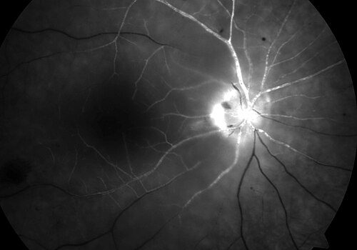
Fluorescein angiography shows delayed arterial filling.
In CRVO, OCT measures macular edema and angiography shows nonperfusion.
So choose imaging by the leading diagnosis.
Systemic Work-Up
CRAO needs carotid imaging, cardiac assessment and inflammatory markers.
CRVO needs blood pressure, glucose and lipid checks.
Younger patients need thrombophilia testing.
Also check intraocular pressure.
CRAO vs CRVO: Treatment of CRAO
No therapy has proved effective in restoring vision.
Ocular massage, paracentesis and thrombolysis have been tried.
Early treatment may help in selected cases (see CRAO management strategies).
Because time matters, refer at once.
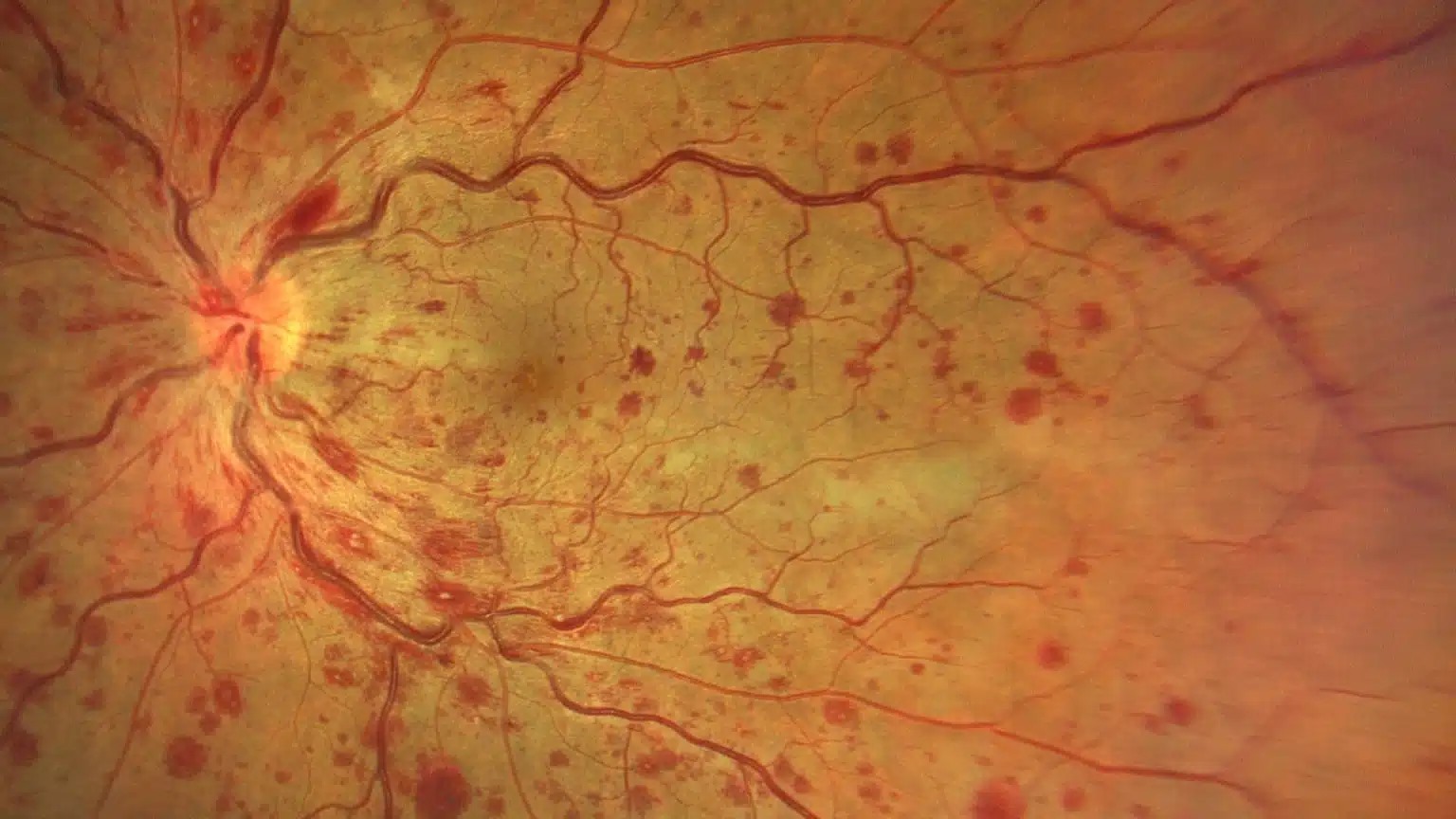
CRAO vs CRVO: Treatment of CRVO
Anti-VEGF injections treat macular edema.
Laser or injections reduce neovascularization in ischemic disease (see anti-VEGF therapy).
Regular follow-up detects neovascular glaucoma.
So treat the complications as well as the occlusion.
CRAO vs CRVO: Complications
CRAO can cause rubeosis and neovascular glaucoma weeks later.
CRVO causes macular edema, neovascularization and glaucoma.
Both need follow-up gonioscopy.
Therefore, watch the iris.
CRAO vs CRVO: Prognosis
Vision in CRAO usually stays poor.
CRVO outcomes vary from full recovery to severe loss.
Early treatment of edema improves the outlook.
So counsel patients on the likely course.
CRAO vs CRVO and Branch Occlusions
Branch retinal artery and vein occlusions affect a sector of the retina (see branch retinal vein occlusion).
The principles are similar but the visual field is only partly lost.
Branch arterial occlusion also needs an embolic work-up.
Also treat edema with injections.
CRAO vs CRVO: Patient Counseling
Explain that artery occlusion is a warning of stroke.
Explain that vein occlusion needs control of blood pressure and glucose.
Both need regular visits.
So give clear information.
CRAO vs CRVO: Practical Summary
Look at the retina, check the pupil and decide the urgency.
If the picture suggests CRAO, send the patient for same-day stroke care.
Because CRVO needs injections, arrange a retina visit.
Finally, review systemic risk factors.
Pitfalls to Avoid
- Do not treat CRAO as a routine retinal problem
- Never forget giant cell arteritis in older patients
- Avoid missing neovascular glaucoma after either occlusion
- Do not skip the systemic work-up
Time Is Critical in Arterial Occlusion
Because retinal tissue tolerates ischemia for only a short time, speed matters.
If the patient arrives within hours, send them to an emergency center.
However, late arrival does not remove the stroke risk.
Therefore, never turn the patient away.
Follow-Up After Occlusion
When the acute phase has passed, review the eye for new vessels.
If the retina is ischemic, treat early.
Because glaucoma can follow, check pressure and the angle.
Finally, coordinate with the physician.
For the latest studies, see the PubMed results for CRAO vs CRVO.


Document what you see
Two smartphone imaging tools built for everyday clinic use — one for the slit lamp, one for the fundus.
From Choroida — the team behind this siteReferences
- Hayreh SS. Ocular vascular occlusive disorders: natural history of visual outcome. Prog Retin Eye Res. 2014;41:1-25.
- Mac Grory B, Schrag M, Biousse V, et al. Management of central retinal artery occlusion: a scientific statement from the American Heart Association. Stroke. 2021;52:e282-e294.
- Central Vein Occlusion Study Group. Natural history and clinical management of central retinal vein occlusion. Arch Ophthalmol. 1997;115:486-491.
- Brown DM, Campochiaro PA, Singh RP, et al. Ranibizumab for macular edema following central retinal vein occlusion: six-month primary end point results of a phase III study. Ophthalmology. 2010;117:1124-1133.
- Rogers S, McIntosh RL, Cheung N, et al. The prevalence of retinal vein occlusion: pooled data from population studies from the United States, Europe, Asia, and Australia. Ophthalmology. 2010;117:313-319.

Writes and reviews the clinical articles on Educate. Every post is checked against peer-reviewed sources before it goes live.