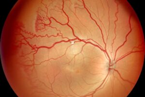
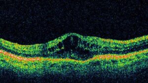
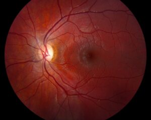
Thrombolysis for central retinal artery occlusion aims to restore flow before the inner retina dies.
Retinal tissue tolerates complete ischemia for only a short time.
Therefore, the question is if thrombolysis works and how early it must start.
Why Thrombolysis for Central Retinal Artery Occlusion Was Tried
Central retinal artery occlusion usually causes severe, permanent vision loss.
The artery is a branch of the ophthalmic artery, so a clot or embolus often causes it.
Also, thrombolysis has proved useful in ischemic stroke from similar emboli.
As a result, clinicians tested it in the eye.
Natural History of the Disease
Most eyes with untreated central retinal artery occlusion end with vision of counting fingers or worse.
A cilioretinal artery can spare the macula in a minority of patients.
Also, Hayreh’s primate work showed that irreversible damage occurs after about 90 to 240 minutes of total occlusion.
Therefore, delays of many hours limit the benefit of any treatment.
Fundus Explorer Pro
Photograph the retinal findings described here with the phone already in your pocket — 22 D optics and built-in illumination in one handheld unit.
From Choroida — the team behind this siteThe EAGLE Trial
EAGLE was a randomized trial of local intra-arterial fibrinolysis against conservative treatment in 84 patients.
It enrolled patients within 20 hours of symptom onset.
Intra-arterial alteplase was given through a catheter in the ophthalmic artery.
In addition, conservative therapy included hemodilution and eyeball massage.
EAGLE Results
Visual outcomes did not differ between the two groups.
Adverse events were more frequent after fibrinolysis, and the trial stopped early.
Also, the window of up to 20 hours was long for retinal tissue.
Therefore, EAGLE did not support routine intra-arterial treatment in late presenters.
Intravenous Thrombolysis for Central Retinal Artery Occlusion
Case series and a patient-level meta-analysis examined intravenous alteplase.
The pooled data suggested a better chance of good vision in patients treated within 4.5 hours.
Benefits faded when treatment started later.
Also, intracranial hemorrhage was uncommon, but data came from nonrandomized reports.
Randomized Trials of Thrombolysis for Central Retinal Artery Occlusion
Newer randomized trials test alteplase or tenecteplase within 4.5 hours.
Their results will define the role of thrombolysis in the first hours.
Also, trial designs match the stroke pathway.
Because of this, follow current guidelines until the data are complete.
Treat Central Retinal Artery Occlusion as Stroke
The American Heart Association now advises managing the condition as an acute ischemic event.
Patients with symptoms for under 4.5 hours should go to a stroke center without delay.
Also, a stroke workup is needed in every patient (see central retinal artery occlusion).
Therefore, the eye clinic should call the emergency system early.
Selecting Patients for Thrombolysis for Central Retinal Artery Occlusion
Consider thrombolysis in patients with sudden painless vision loss and a clear time of onset.
The fundus shows a cherry-red spot and retinal whitening.
Also, exclude giant cell arteritis in older patients, since steroids are the urgent treatment there.
In addition, review contraindications to thrombolysis.
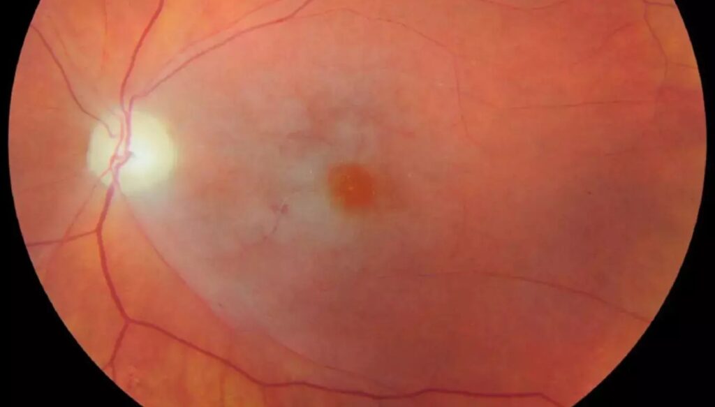
Contraindications to Thrombolysis for Central Retinal Artery Occlusion
Recent major surgery, active bleeding and uncontrolled hypertension are the main ones.
A history of intracranial hemorrhage is also a barrier.
Also, an unclear time of onset limits the use.
Because of this, the decision belongs to a stroke team.
Conservative Measures
Ocular massage, anterior chamber paracentesis and carbogen have no proven benefit.
They carry little risk and may be tried when nothing else is available.
Also, lowering intraocular pressure rarely changes the outcome.
Therefore, do not delay transfer for these maneuvers.
Systemic Workup
Every patient needs carotid imaging, cardiac evaluation and blood tests.
Inflammatory markers help detect giant cell arteritis.
Also, atrial fibrillation and carotid disease are the most common sources.
In addition, start secondary prevention at once.
What to Tell Patients
Explain that vision may not return, even with treatment.
The same event raises the risk of stroke and heart disease in the following weeks.
Also, urgent systemic care protects life more than vision.
Therefore, counsel patients early and honestly.
Pitfalls to Avoid
- Do not wait for a retina specialist before sending the patient to a stroke center
- Never give thrombolysis outside a stroke pathway
- Do not forget to rule out giant cell arteritis
- Never rely on ocular massage as definitive treatment
The Time Window
The 4.5-hour window comes from stroke trials.
Retinal tissue is probably more vulnerable than brain tissue.
Also, delays at triage waste precious minutes.
Therefore, speed matters at every step.
Role of the Ophthalmologist
Confirm the diagnosis with a dilated fundus examination.
Record the exact time of onset and call the stroke team.
Also, avoid tests that delay transfer.
As a result, the patient reaches treatment sooner.


Document what you see
Two smartphone imaging tools built for everyday clinic use — one for the slit lamp, one for the fundus.
From Choroida — the team behind this siteReferences
- Schumacher M, Schmidt D, Jurklies B, et al. Central retinal artery occlusion: local intra-arterial fibrinolysis versus conservative treatment, a multicenter randomized trial. Ophthalmology. 2010;117:1367-1375.
- Schrag M, Youn T, Schindler J, Kirshner H, Greer D. Intravenous fibrinolytic therapy in central retinal artery occlusion: a patient-level meta-analysis. JAMA Neurol. 2015;72:1148-1154.
- Mac Grory B, Schrag M, Biousse V, et al. Management of central retinal artery occlusion: a scientific statement from the American Heart Association. Stroke. 2021;52:e282-e294.
- Hayreh SS, Zimmerman MB. Central retinal artery occlusion: visual outcome. Am J Ophthalmol. 2005;140:376-391.

Writes and reviews the clinical articles on Educate. Every post is checked against peer-reviewed sources before it goes live.