Corneal perforation is a full-thickness defect in the cornea, and it needs prompt treatment to avoid loss of the eye.
It follows ulceration from infection, melting from autoimmune disease, trauma, or chemical injury.
In addition, small perforations can be sealed with cyanoacrylate glue, and larger ones need a graft.
What Happens in a Corneal Perforation?
When the stroma melts or is lost, Descemet membrane may bulge as a descemetocele, and then give way (see descemetocele).
Aqueous humor leaks out, the anterior chamber becomes shallow or flat, and the iris may also plug the hole.
The eye is at risk of infection, synechiae, cataract, and glaucoma, so the aim is to restore the integrity of the globe and treat the cause.
Common Causes
- Infectious keratitis, bacterial and fungal (see fungal versus bacterial keratitis)
- Sterile melting, with rheumatoid arthritis and other collagen vascular diseases (see corneal melt and peripheral ulcerative keratitis)
- Neurotrophic and exposure keratopathy (see neurotrophic keratitis)
- Herpetic keratitis and chemical burns (see chemical eye injury)
- Trauma and surgical wound problems (see corneal wound dehiscence)
All-fit Slit-Lamp Adapter
Record and share exactly what you see at the slit lamp. One adapter fits any slit lamp or surgical microscope — and any smartphone.
From Choroida — the team behind this siteDiagnosis of Corneal Perforation
Check for a shallow chamber, an iris plug, and a leaking wound, and perform a Seidel test with fluorescein.
In a positive test, a stream of clear aqueous dilutes the dye, and this is also a sign of an active leak.
Also, measure the size of the perforation and the thickness of the surrounding cornea, and take cultures of infectious ulcers.
Avoid applying pressure to the globe, and protect it with a shield.
Cyanoacrylate Tissue Adhesive
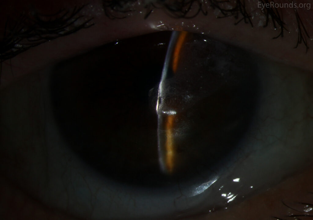
Cyanoacrylate glue polymerizes on contact with moisture, and it forms a hard plug that seals a small hole and provides temporary support.
It is also suitable for perforations of up to about 2 to 3 millimeters, with a chamber that can be formed, and a thin ulcer bed.
Leahey and colleagues reported success with cyanoacrylate for severe corneal thinning and perforation, and the technique is a standard first-line measure for small perforations.
Technique of Corneal Perforation
- Clean the area and remove loose epithelium and necrotic tissue around the defect
- Dry the area, as the glue should be applied on a dry surface
- Place a small drop of glue on the tip of an applicator, such as a sterile plastic or a fine disc, and apply it to the defect, or apply directly with a fine tip
- Allow the glue to polymerize for a minute, forming a thin film
- Cover with a bandage contact lens to protect the glue and the eye
- Check for leakage, and form the anterior chamber with viscoelastic or air if it is flat
A thin layer is better than a thick one, which is more irritating and may dislodge.
After Gluing
- Topical antibiotics and cycloplegics, with aqueous suppressants to lower the pressure
- Treatment of the underlying disease, including infection, inflammation, and dry eye
- Careful follow-up, with checks for leakage, glue position, and infection
- Avoid eye rubbing and strain, and consider an eye shield
The glue may stay in place for weeks to months, and it typically falls off as the cornea heals underneath, or it can be replaced.
Larger Perforations and Alternatives
- Amniotic membrane and multilayered grafts for small to moderate defects
- Tectonic patch graft, a full-thickness or lamellar corneal graft that fills the defect and restores integrity
- Therapeutic penetrating or lamellar keratoplasty, for larger or infected perforations (see penetrating keratoplasty and deep anterior lamellar keratoplasty)
- Conjunctival flap, a temporary option in difficult eyes
Hirst and colleagues reviewed the changing treatments for corneal perforation, and Jhanji and colleagues published a modern review of management.
Corneal Perforation: Complications and Counselling
Glue can cause inflammation, vascularization, and cataract if it contacts the lens, and it may fail to hold in large or leaking defects.
Explain to the patient that the glue is a temporary measure that buys time, that a graft may still be needed, and that vision may be limited by scarring.
Advise immediate return if there is pain, redness, or sudden change in vision.
Practical Notes
Keep glue and applicators ready in the emergency room, since the time between the diagnosis and the treatment is important.
Also, use a sterile technique and sterile glue, and use a fresh applicator for each patient.
Avoid getting glue on the eyelids or the skin, and keep the eye still during the polymerization.
If the glue is dislodged, repeat the procedure after drying the area.
Follow-Up Plan
Review the patient frequently in the first week, and check for leaks, infection, and the position of the glue.
Continue the treatment of the cause, and plan definitive repair if the defect is large or the underlying disease is progressive.
In addition, document the date of gluing and the plan for follow-up in the chart.
For the latest studies, see the PubMed results for corneal perforation.


Document what you see
Two smartphone imaging tools built for everyday clinic use — one for the slit lamp, one for the fundus.
From Choroida — the team behind this siteReferences
- Leahey AB, Gottsch JD, Stark WJ. Clinical experience with N-butyl cyanoacrylate (Nexacryl) tissue adhesive. Ophthalmology. 1993;100:173-180.
- Hirst LW, Smiddy WE, Stark WJ. Corneal perforations: changing methods of treatment, 1960-1980. Ophthalmology. 1982;89:630-635.
- Jhanji V, Young AL, Mehta JS, Sharma N, Agarwal T, Vajpayee RB. Management of corneal perforation. Surv Ophthalmol. 2011;56:522-538.
- American Academy of Ophthalmology. Basic and Clinical Science Course, Section 8: External Disease and Cornea. AAO; current edition.
Test yourself
A few questions straight from this article.
-
As the stroma melts toward a corneal perforation, what may form before Descemet membrane gives way?
When the stroma melts or is lost, Descemet membrane may bulge as a descemetocele and then give way, producing a full-thickness defect. -
In a suspected corneal perforation, what does a positive Seidel test with fluorescein show?
In a positive Seidel test a stream of clear aqueous dilutes the fluorescein, a sign of an active leak. -
Which examination findings point to a corneal perforation?
Aqueous leaks out, so the chamber becomes shallow or flat, and the iris may plug the hole; check also for a leaking wound. -
Up to roughly what size of corneal perforation is cyanoacrylate glue suitable?
Glue suits perforations up to about 2 to 3 millimeters with a formable chamber and a thin ulcer bed; larger defects need a graft. -
How does cyanoacrylate tissue adhesive seal a small corneal perforation?
Cyanoacrylate polymerizes on contact with moisture, forming a hard plug that seals a small hole and gives temporary support. -
Why is a thin layer of cyanoacrylate glue preferred over a thick one for corneal perforation?
A thin film is better because a thick layer is more irritating and more likely to dislodge. -
After cyanoacrylate gluing of a corneal perforation, what is placed to protect the glue and the eye?
The glue is covered with a bandage contact lens; pressure on the globe is avoided and a shield can be used. -
Which treatment is used for a larger or infected corneal perforation?
Larger or infected perforations need therapeutic penetrating or lamellar keratoplasty; a tectonic patch graft can also fill the defect and restore integrity. -
Which complication can cyanoacrylate glue cause in corneal perforation if it contacts the lens?
Glue can cause inflammation and vascularization, and cataract if it contacts the lens; it may also fail to hold in large or leaking defects. -
How long does cyanoacrylate glue typically remain on a sealed corneal perforation?
The glue may stay for weeks to months and typically falls off as the cornea heals beneath it, or it can be replaced; it is a temporary measure that buys time.

Writes and reviews the clinical articles on Educate. Every post is checked against peer-reviewed sources before it goes live.