
An optic disc coloboma traces back to a single, specific embryologic event, the failure of the fetal (choroidal) fissure to close completely during early gestation, and this shared origin is why coloboma so often involves more than just the optic disc in isolation, extending to the iris, choroid, and retina along the same embryologic line, and why its presence should always prompt a broader look for both ocular and systemic associations.
The Embryology Behind the Defect
During normal eye development, a groove called the fetal fissure runs along the underside of the developing optic cup and stalk, allowing blood vessels to enter the eye during this critical period, and this fissure normally closes completely by around the sixth to seventh week of gestation.
When closure is incomplete, a coloboma results, and depending on exactly where along this fissure closure failed, the resulting defect can involve the iris, ciliary body, choroid, retina, and optic disc in various combinations, which is why a disc coloboma is often, though not always, found alongside coloboma involving these other, embryologically related structures.
Clinical Appearance
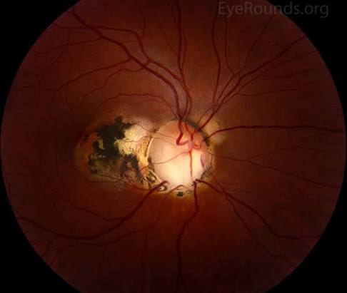
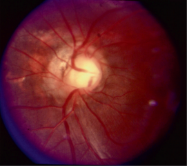
- A characteristic, often large, excavated, bowl-shaped or glistening white defect involving the optic disc, typically most pronounced inferiorly, reflecting the inferior location of the fetal fissure during normal development
- The disc is usually enlarged compared with normal, and the excavation can extend into the adjacent peripapillary retina and choroid
- Often associated with a visible, pale, thinned or absent retina and choroid overlying the excavated area, given the same developmental defect affecting these embryologically related tissues
- Can be unilateral or bilateral, and the severity and extent of involvement vary considerably between affected individuals
Fundus Explorer Pro
Photograph the retinal findings described here with the phone already in your pocket — 22 D optics and built-in illumination in one handheld unit.
From Choroida — the team behind this siteVisual Consequences
- Visual acuity can range from normal to severely impaired, depending heavily on whether the coloboma involves the papillomacular bundle or extends to affect the macula directly
- A visual field defect, often a superior field defect corresponding to the inferior anatomic location of the excavated disc and retina, is common
- Strabismus and, in more severe or asymmetric bilateral cases, nystagmus can develop related to poor or asymmetric vision
Systemic Associations
Optic disc coloboma, particularly when bilateral or accompanied by other ocular coloboma (iris, choroid), should prompt evaluation for associated systemic conditions, since coloboma is a recognized component of several syndromes.
- CHARGE syndrome, an important association to specifically consider, given its combination of coloboma, heart defects, choanal atresia, growth and developmental delay, genital abnormalities, and ear anomalies, several of which carry significant, sometimes urgent, management implications
- Other chromosomal and genetic syndromes have also been associated with optic disc and other ocular coloboma, supporting a broader genetic evaluation in appropriate clinical contexts
Retinal Detachment Risk
A meaningful proportion of eyes with optic disc coloboma, particularly when the coloboma extends to involve the adjacent retina, carry an elevated lifetime risk of retinal detachment, related to the abnormal, often thinned and poorly adherent retinal tissue at the margin of the coloboma, which is why ongoing surveillance for this specific complication is an important part of long-term follow-up in these patients.
Diagnostic Evaluation
- Dilated fundus examination characterizing the extent and location of the disc excavation and any associated choroidal or retinal involvement
- Careful examination of the iris and anterior segment for any associated coloboma in these structures
- Visual field testing, when age and cooperation allow, to characterize the functional visual impact
- Systemic evaluation, including cardiac, renal, and hearing assessment, and genetic evaluation when the clinical picture suggests an associated syndrome, particularly CHARGE syndrome
- Optical coherence tomography can help characterize the detailed structure of the coloboma and any subtle associated retinal changes not fully appreciated on direct examination alone
Management
Vision-Related Management
- Correction of any associated refractive error and management of amblyopia risk, particularly important in unilateral or asymmetric bilateral cases, applying standard amblyopia treatment principles during the critical period of visual development
- Low vision support for patients with significant visual field or acuity impairment from extensive coloboma involvement
Monitoring for Retinal Detachment
Given the elevated lifetime risk, patients with optic disc coloboma involving adjacent retina benefit from patient education about the symptoms of retinal detachment and appropriately regular dilated fundus examination to monitor for this complication over time.
Coordinated Systemic Care
When an associated systemic syndrome, particularly CHARGE syndrome, is identified or suspected, coordinated multidisciplinary care addressing the full range of associated systemic findings is essential, given the potentially serious implications of some of these associated features beyond the eye itself.
Prognosis
Visual prognosis in optic disc coloboma varies enormously depending on the extent of disc and macular involvement, ranging from good, functional vision in milder, more peripherally confined cases to significant visual impairment when the papillomacular bundle or macula itself is affected.
Beyond the visual prognosis, recognizing the potential for associated systemic syndromes, and the lifelong retinal detachment risk in affected eyes, supports the broader, coordinated, and appropriately sustained care this congenital condition requires.


Document what you see
Two smartphone imaging tools built for everyday clinic use — one for the slit lamp, one for the fundus.
From Choroida — the team behind this siteReferences
- Onwochei BC, Simon JW, Bateman JB, Couture KC, Mir E. Ocular colobomata. Surv Ophthalmol. 2000;45:175-194.
- Pagon RA. Ocular coloboma. Surv Ophthalmol. 1981;25:223-236.
- Blake KD, Prasad C. CHARGE syndrome. Orphanet J Rare Dis. 2006;1:34.
- Gregory-Evans CY, Williams MJ, Halford S, Gregory-Evans K. Ocular coloboma: a reassessment in the age of molecular neuroscience. J Med Genet. 2004;41:881-891.
