
Conjunctival amyloidosis produces a waxy, often vascularized subconjunctival mass that can look concerning enough to prompt biopsy on appearance alone, and the biopsy that follows serves two purposes at once: confirming the diagnosis and, just as importantly, prompting the question every finding of amyloid deposition must answer, whether this is a purely localized, harmless process or a visible sign of systemic amyloidosis affecting other organs.
What Amyloid Is Doing in the Conjunctiva
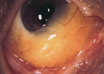
Amyloid is an abnormal, misfolded protein that can deposit in tissue either as a localized, isolated phenomenon or as part of a systemic disease process affecting multiple organs, and conjunctival amyloidosis most commonly represents primary localized amyloidosis, confined to the conjunctiva itself without any associated systemic disease, though the possibility of systemic involvement must always be actively considered and excluded rather than assumed.
Clinical Presentation
- A slowly enlarging, waxy, yellow to pink-white subconjunctival mass or diffuse thickening, most often located in the fornix or bulbar conjunctiva
- Prominent, often tortuous blood vessels coursing over or within the lesion, since amyloid deposition can weaken vessel walls and lead to increased vascular fragility
- Recurrent spontaneous subconjunctival hemorrhage is a characteristic and sometimes presenting feature, related to this vascular fragility, and recurrent unexplained subconjunctival bleeding in the same location should raise suspicion for an underlying amyloid deposit rather than being dismissed as simple recurrent benign hemorrhage
- Generally slow-growing over months to years, and can cause mechanical irritation or cosmetic concern as it enlarges
All-fit Slit-Lamp Adapter
Record and share exactly what you see at the slit lamp. One adapter fits any slit lamp or surgical microscope — and any smartphone.
From Choroida — the team behind this siteDiagnostic Evaluation
Biopsy
Biopsy with histopathologic examination is required for diagnosis, with special staining, most classically Congo red staining demonstrating the characteristic apple-green birefringence under polarized light, confirming the presence of amyloid material within the tissue.
Systemic Evaluation
Once conjunctival amyloidosis is confirmed histologically, systemic evaluation is an essential next step to distinguish primary localized disease, confined to the conjunctiva alone, from a manifestation of systemic amyloidosis, which can affect the heart, kidneys, nervous system, and other organs with serious implications.
- Evaluation for systemic symptoms and signs suggestive of amyloid involvement elsewhere, including unexplained proteinuria, cardiac dysfunction, or peripheral neuropathy
- Laboratory testing, including serum and urine protein electrophoresis, when systemic amyloidosis, particularly the form associated with plasma cell disorders, is a concern
- Coordination with hematology or internal medicine for more complete systemic evaluation when any suggestive findings are present
Management
Localized Disease
For confirmed primary localized conjunctival amyloidosis without evidence of systemic involvement, surgical excision of the mass is the standard treatment for symptomatic, enlarging, or cosmetically bothersome lesions, though recurrence at the same site is possible given the chronic, ongoing nature of local amyloid production even after gross excision.
Managing Recurrent Hemorrhage
For lesions causing recurrent, bothersome subconjunctival hemorrhage, surgical excision can also help reduce the frequency of these episodes by removing the fragile, amyloid-affected vascular tissue directly.
Systemic Disease
When systemic amyloidosis is identified, management is directed by the underlying systemic disease and its specific type, coordinated with the appropriate specialists, with the conjunctival lesion itself typically managed locally as needed for symptoms while systemic treatment addresses the broader disease process.
Follow-Up
Even after apparently successful local excision, periodic follow-up is appropriate given the possibility of local recurrence, and any patient with primary localized disease at diagnosis should remain aware that new or evolving systemic symptoms in the future would warrant renewed systemic evaluation, since localized disease at one point in time does not permanently exclude the later development of systemic involvement.
Prognosis
Primary localized conjunctival amyloidosis, once confirmed to be isolated without systemic involvement, has an excellent prognosis, with local excision generally providing good symptomatic and cosmetic results, even accounting for the possibility of local recurrence over time.
The prognosis for systemic amyloidosis, when identified, depends entirely on the specific type and extent of systemic organ involvement, underscoring why the biopsy-confirmed diagnosis of conjunctival amyloidosis should always be followed by a deliberate, systematic assessment for systemic disease rather than assumed to be an isolated ocular curiosity.


Document what you see
Two smartphone imaging tools built for everyday clinic use — one for the slit lamp, one for the fundus.
From Choroida — the team behind this siteReferences
- Leibovitch I, Selva D, Goldberg RA, et al. Periocular and orbital amyloidosis: clinical characteristics, management, and outcome. Ophthalmology. 2006;113:1657-1664.
- Rodman HI, Hardy JH, Barr JW. Localized conjunctival amyloidosis. Am J Ophthalmol. 1968;65:892-899.
- Fahmy JA, Bock J. Conjunctival amyloidosis. Acta Ophthalmol (Copenh). 1976;54:445-452.
- Knowles DM 2nd, Jakobiec FA, Rosen M, Howard G. Amyloidosis of the orbit and adnexae. Surv Ophthalmol. 1975;19:367-384.
