

DISEASE
Bardet-Biedl Syndrome (BBS) is a rare, autosomal recessive genetic disorder that can lead to the dysfunction of multiple organ systems, including the kidneys, genitalia, brain, and eye.

Bardet-Biedl Syndrome (BBS) is caused by pathogenic mutations in genes encoding proteins involved in the function of non-motile primary cilia. Rod and cone photoreceptors do not contain primary cilia but rather have a primary cilia-like structure that spans the inner and outer segments.
Photoreceptor outer segments have therefore been conceptualized as specialized sensory cilia, sometimes referred to as photoreceptor sensory cilia (PSC).
Primary cilia and photoreceptor cilia are structurally similar, are composed of many of the same proteins, and are both dysfunctional in BBS. Proteins affected by BBS are involved in intracellular protein trafficking along the photoreceptor connecting cilium, a process known as intraflagellar transport.
In Bardet-Biedl Syndrome (BBS) mutant photoreceptors, proteins are mislocalized to incorrect cellular substructures. For example, rhodopsin accumulates in rod inner segments and cell bodies at the expense of its localization to outer segments in some BBS mouse models.
Other work shows an abnormal accumulation of 138 proteins in photoreceptor outer segments of BBS mutant mice compared to wild types. The mislocalization and ectopic accumulation of proteins are thought to lead to inadequate cellular homeostasis and ultimately photoreceptor cell death.
Diagnostic procedures
Electroretinography (ERG) testing can support signs of retinal dysfunction, particularly in early cases that have not yet manifested signs on the retinal exam.
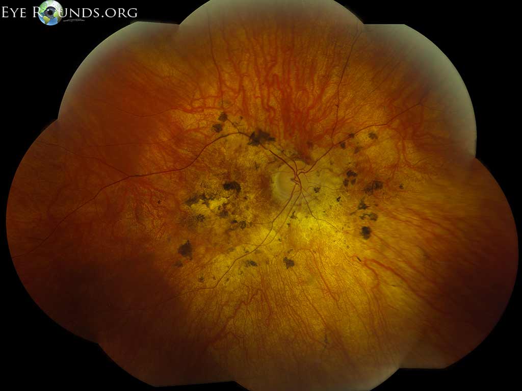
Typically, ERG in someone with BBS shows a mixed rod-cone dystrophy, manifesting as diminished a-waves and b-waves at both scotopic and photopic light levels.
Interestingly, asymptomatic carriers of BBS can also exhibit abnormalities on flash and multifocal ERG testing.
MANAGEMENT
General treatment
There is no therapy to treat the cause of Bardet-Biedl Syndrome (BBS), but multidisciplinary care is required to treat disease manifestations. If present, diabetes, hypertension, and metabolic syndrome are managed intensively to minimize damage to other organ systems also involved in BBS, such as the kidney and retina.
Disease-specific manifestations are managed by respective specialists. Kidney dysfunction is an especially common source of morbidity and mortality in BBS patients.
Obesity and hyperphagia in BBS are hypothesized to be a result of hypothalamic dysfunction. In 2020, treatment with Setmelanotide, an agonist to the melanocortin-4-receptor (MC4R), which regulates appetite and body weight, was shown to result in significant weight loss in patients with BSS over a 1-year period.
This drug continues to demonstrate positive outcomes in patients with BBS.
Visual dysfunction is often significant in BBS. Initial ophthalmological evaluation in children should include assessment for strabismus, nystagmus, and decreased visual acuity, and if the patient is mature enough, electroretinography and visual field testing.
Patients should be referred for low-vision services as indicated. Annual or more frequent follow-up, as needed, with an ophthalmologist is recommended.
Surgery
Surgical intervention may be required for anatomic abnormalities, such as orodental, cardiovascular, and genitourinary malformations, but there is currently no role for surgery for retinal degeneration in BBS.
Would you have interest in taking retinal images with your smartphone?
Fundus photography is superior to fundus analysis as it enables intraocular pathologies to be photo-captured and encrypted information to be shared with colleagues and patients.
Recent technologies allow smartphone-based attachments and integrated lens adaptors to transform the smartphone into a portable fundus camera and Retinal imaging by smartphone.
RETINAL IMAGING BY YOUR SMARTPHONE
REFERENCES
- Forsyth R, Gunay-Aygun M. Bardet-Biedl Syndrome Overview. 2003 Jul 14 [updated 2020 Jul 23]. In: Adam MP, Ardinger HH, Pagon RA, Wallace SE, Bean LJH, Gripp KW, Mirzaa GM, Amemiya A, editors. GeneReviews®. Seattle (WA): University of Washington, Seattle; 1993–2021. PMID: 20301537.
- Forsythe, E., & Beales, P. L. (2013). Bardet-Biedl syndrome. European Journal of Human Genetics, 21(1), 8–13. https://doi.org/10.1038/ejhg.2012.115.
- Forsythe, E., Kenny, J., Bacchelli, C., & Beales, P. L. (2018). Managing Bardet-Biedl Syndrome-Now and in the Future. Frontiers in Pediatrics, 6, 23. https://doi.org/10.3389/fped.2018.00023.
- Weihbrecht, K. (2020). Bardet-Biedl syndrome. In Genetics and Genomics of Eye Disease (pp. 117–136). Elsevier. https://doi.org/10.1016/B978-0-12-816222-4.00008-3.
- Bujakowska, Kinga M., Qin Liu, and Eric A. Pierce. “Photoreceptor Cilia and Retinal Ciliopathies.” Cold Spring Harbor Perspectives in Biology 9, no. 10 (October 3, 2017). https://doi.org/10.1101/cshperspect.a028274.
- Weihbrecht, K., Goar, W. A., Pak, T., Garrison, J. E., DeLuca, A. P., Stone, E. M., Scheetz, T. E., & Sheffield, V. C. (2017). Keeping an Eye on Bardet-Biedl Syndrome: A Comprehensive Review of the Role of Bardet-Biedl Syndrome Genes in the Eye. Medical Research Archives, 5(9). https://doi.org/10.18103/mra.v5i9.1526.
