
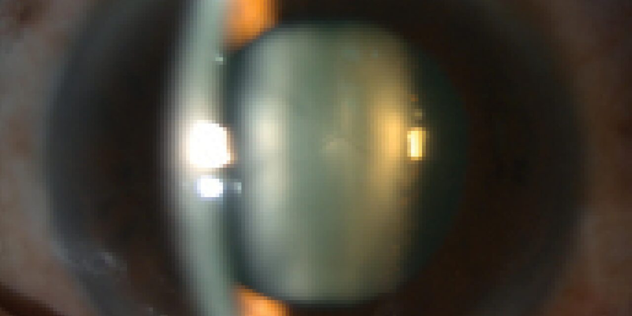
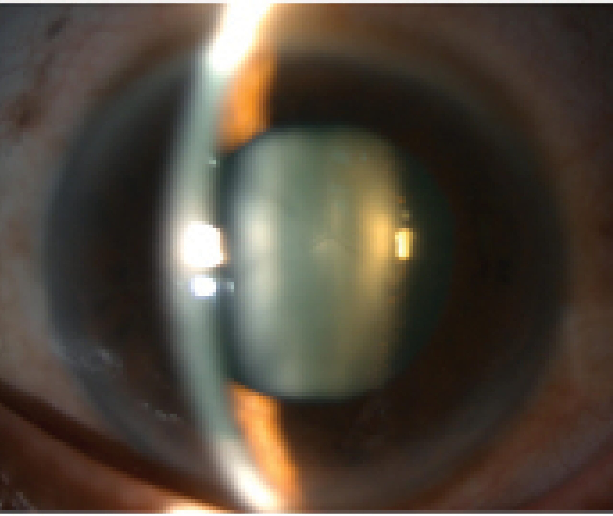
Lens subluxation is diagnosed at the slit lamp through a set of specific, learnable signs, phacodonesis, iridodonesis, and asymmetric anterior chamber depth chief among them, and picking up on these subtle clues during a routine examination can be the first step toward uncovering an underlying systemic connective tissue disorder the patient did not know they had.
What Subluxation Means
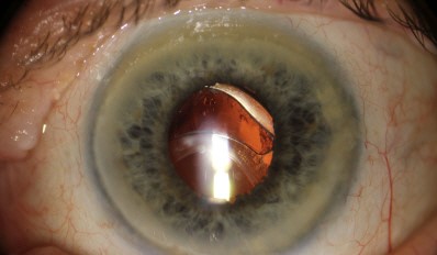
Lens subluxation refers to partial displacement of the lens from its normal centered position, resulting from weakness, stretching, or focal breakdown of the zonular fibers that normally hold the lens securely centered behind the iris, distinguished from complete lens dislocation, in which the zonular support has failed entirely and the lens has moved fully out of the pupillary space, often into the anterior chamber or vitreous cavity.
Key Clinical Signs
Phacodonesis
A trembling or wobbling movement of the lens itself, best observed at the slit lamp as the patient makes a saccadic eye movement and then stops, with the lens visibly continuing to oscillate briefly afterward due to its inadequate zonular anchoring, a direct and often the most reliable sign of zonular weakness.
Iridodonesis
A similar trembling movement of the iris, occurring for the same underlying reason, since the iris normally rests against and is partly supported by the anterior lens surface, so a poorly anchored lens allows the iris to move more than normal with eye movement as well.
Asymmetric or Abnormal Anterior Chamber Depth
A subluxated lens tilted or displaced within the eye can produce localized shallowing or deepening of the anterior chamber depending on the direction of displacement, sometimes asymmetric between different clock-hour positions around the same eye, a subtler but valuable sign on careful examination.
Visible Lens Edge
In more significant subluxation, the edge of the lens itself, and sometimes visible zonular fibers, can become apparent within the pupillary space, particularly with pupillary dilation, directly confirming the diagnosis.
All-fit Slit-Lamp Adapter
Record and share exactly what you see at the slit lamp. One adapter fits any slit lamp or surgical microscope — and any smartphone.
From Choroida — the team behind this siteCauses
- Marfan syndrome, classically causing bilateral, often symmetric, superotemporal lens subluxation, related to an underlying fibrillin connective tissue abnormality affecting the zonular fibers
- Homocystinuria, another connective tissue-related metabolic disorder, classically causing inferonasal lens subluxation, a directional pattern that helps distinguish it clinically from Marfan-related subluxation
- Trauma, causing focal zonular dialysis (tearing) at the site of impact, producing asymmetric, often unilateral subluxation in the direction opposite the traumatic force
- Pseudoexfoliation syndrome, a common cause of more generalized, gradual zonular weakness, particularly relevant in cataract surgery planning given its associated intraoperative risk (see zonular dehiscence in cataract surgery)
- Ectopia lentis et pupillae and other less common genetic causes, each with their own characteristic patterns (see ectopia lentis et pupillae)
Diagnostic Evaluation
- Careful dynamic observation for phacodonesis and iridodonesis during eye movement, one of the more revealing parts of the slit-lamp examination for this diagnosis
- Assessment of anterior chamber depth and any asymmetry around the circumference of the angle
- A directed history for trauma, and a family and personal history and systemic examination for features of Marfan syndrome or homocystinuria when the pattern or bilaterality of subluxation suggests a genetic cause
- Gonioscopy and, when needed, ultrasound biomicroscopy to further characterize the extent and location of zonular loss, particularly useful for surgical planning
Clinical and Surgical Implications
Recognizing lens subluxation before cataract surgery is undertaken is essential, since it fundamentally changes the surgical approach and risk profile, prompting consideration of capsular support devices, modified surgical technique, and, for more severe subluxation, alternative intraocular lens fixation strategies (see capsular tension ring indications).
Missing this diagnosis preoperatively and proceeding with standard cataract surgery technique significantly increases the risk of intraoperative complications, including posterior capsular rupture and lens fragment dislocation.
Prognosis
With correct preoperative recognition of lens subluxation and appropriate surgical planning, good visual outcomes are achievable even in eyes with significant zonular compromise.
Beyond the immediate ocular management, identifying lens subluxation, particularly bilateral subluxation with a directional pattern suggesting Marfan syndrome or homocystinuria, carries broader diagnostic value, since these underlying systemic conditions carry their own serious health implications warranting appropriate further evaluation and management.


Document what you see
Two smartphone imaging tools built for everyday clinic use — one for the slit lamp, one for the fundus.
From Choroida — the team behind this siteReferences
- Nelson LB, Maumenee IH. Ectopia lentis. Surv Ophthalmol. 1982;27:143-160.
- Cross HE, Jensen AD. Ocular manifestations in the Marfan syndrome and homocystinuria. Am J Ophthalmol. 1973;75:405-420.
- Jarrett WH 2nd. Dislocation of the lens: a study of 166 hospitalized cases. Arch Ophthalmol. 1967;78:289-296.
- Halpert M, BenEzra D. Surgery of the hereditary subluxated lens in children. Ophthalmology. 1996;103:681-686.
