
Photokeratitis is a painful, acute inflammation of the corneal epithelium caused by excessive ultraviolet radiation exposure — a sunburn of the cornea.
It produces one of the more dramatic mismatches in ophthalmology between how minor the exposure often felt at the time and how severe the resulting pain turns out to be several hours later.
The delay between exposure and symptom onset is itself a diagnostic clue, because it distinguishes photokeratitis from most other acute causes of a painful red eye that produce symptoms immediately.
Common Scenarios
- Welding arc exposure without proper eye protection (“arc eye,” “welder’s flash”) — one of the most common occupational causes
- Snow blindness — UV reflection off snow at altitude, particularly in mountaineering and skiing without adequate UV-blocking eyewear
- Tanning bed use without appropriate goggles
- Direct or indirect solar viewing, including during a solar eclipse without proper filtration (though this mechanism, along with true solar retinopathy, involves different, often deeper retinal injury discussed in this site’s separate coverage of solar retinopathy)
- Germicidal UV lamp exposure, including from improperly used UV sanitizing devices
- High-altitude sun exposure without adequate eye protection, from the combination of thinner atmosphere and reflection off snow or water
Pathogenesis
UV-B radiation is absorbed primarily by the corneal epithelium, where it damages epithelial cell DNA and membranes, triggering apoptosis and shedding of the superficial epithelial layer.
The characteristic delay of several hours between exposure and symptom onset reflects the time needed for this epithelial cell death and desquamation to actually manifest clinically.
The damage is done at the time of exposure, but the clinical picture develops afterward as the affected cells die and are shed.
All-fit Slit-Lamp Adapter
Record and share exactly what you see at the slit lamp. One adapter fits any slit lamp or surgical microscope — and any smartphone.
From Choroida — the team behind this siteClinical Presentation
Patients typically present hours after UV exposure with sudden, severe bilateral eye pain, a sensation often described as intensely gritty or as if sand were in the eyes, along with photophobia, tearing, blepharospasm, and significant difficulty keeping the eyes open.
The severity of pain relative to what is often, in retrospect, described as a relatively brief exposure can be striking, and patients frequently present to urgent or emergency care given how acutely disabling the symptoms are.
Exam Findings
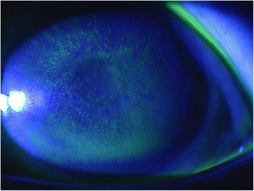
- Diffuse, fine, punctate epithelial defects across the cornea, typically most dense in the interpalpebral zone corresponding to the area of UV exposure not covered by the eyelids
- Conjunctival injection
- Marked photophobia and blepharospasm, sometimes severe enough to make examination difficult without topical anesthesia
- Normal anterior chamber and intraocular pressure, without the deeper findings seen in more serious causes of acute painful red eye
- Bilateral involvement in most cases, reflecting the typically symmetric nature of the exposure
Differential Diagnosis
- Recurrent corneal erosion — usually unilateral, often with a history of prior corneal abrasion, and without the characteristic UV exposure history
- Chemical or thermal burn — a specific exposure history to a chemical agent or heat source rather than light, with potentially deeper tissue involvement depending on severity
- Contact lens overwear or hypoxia — related to lens wear history rather than UV exposure, and typically without the same acute, severe pain onset pattern
- Herpetic keratitis — usually unilateral, with a dendritic pattern on fluorescein staining rather than diffuse fine punctate staining
The history is usually definitive: a clear exposure to welding, snow, tanning beds, or another significant UV source in the preceding several hours, combined with bilateral, symmetric findings, usually makes photokeratitis straightforward to diagnose without further testing.
Management
Photokeratitis is self-limited, and the corneal epithelium typically heals completely within 24 to 48 hours given its excellent regenerative capacity, with management focused entirely on comfort during that window rather than on altering the underlying healing process.
Preservative-free lubricating drops and ointment, oral analgesics, and, for severe pain, a short course of cycloplegia to relieve ciliary spasm all help manage symptoms.
Topical anesthetic drops, meanwhile, are avoided for repeated patient use outside the clinical exam, because they impair epithelial healing and can mask worsening symptoms if used repeatedly at home.
A bandage contact lens can improve comfort in more severe cases by protecting the healing epithelium from the mechanical friction of blinking, though most cases resolve well with simple supportive measures alone.
Given the excellent prognosis and rapid healing, patients can generally be reassured that full recovery without lasting visual consequence is the expected course, and the encounter is a good opportunity for counseling on appropriate UV-protective eyewear to prevent recurrence.


Document what you see
Two smartphone imaging tools built for everyday clinic use — one for the slit lamp, one for the fundus.
From Choroida — the team behind this siteReferences
- Cullen AP. Photokeratitis and other phototoxic effects on the cornea and conjunctiva. International Journal of Toxicology.
- Bergmanson JP, Sheldon TM. Ultraviolet radiation revisited. CLAO Journal.
- American Academy of Ophthalmology. Basic and Clinical Science Course, Section 8: External Disease and Cornea.
- Bagheri N, Wajda B, eds. The Wills Eye Manual.
