
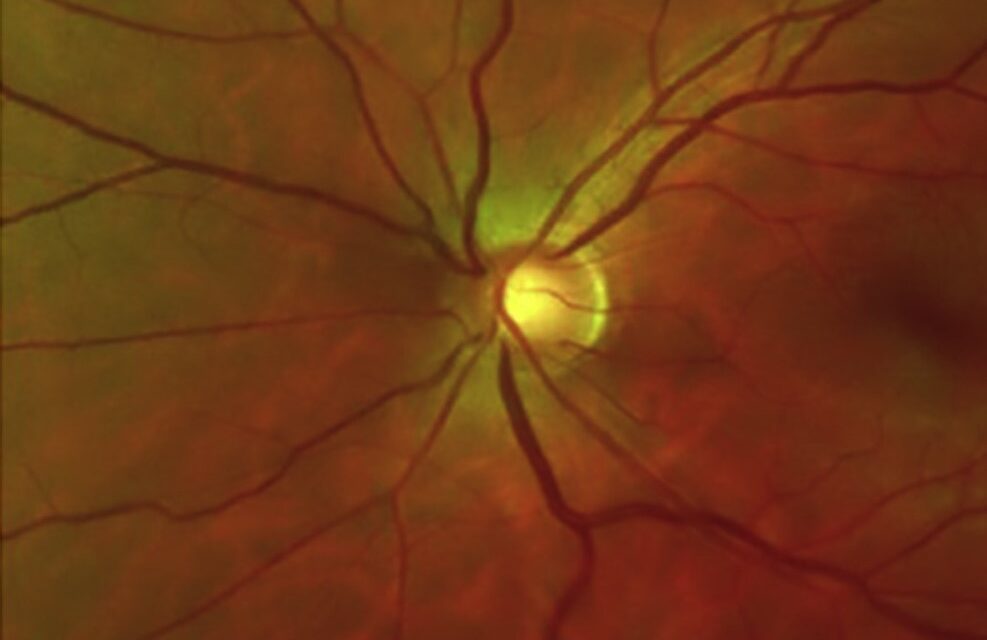
Schwartz-Matsuo Syndrome is a trap that punishes anyone who treats the pressure and forgets to look at the retina.
The eye has a raised intraocular pressure and cells floating in the anterior chamber, so it looks for all the world like inflammatory glaucoma.
Yet the real problem is at the back: a rhegmatogenous retinal detachment is quietly driving the pressure up.
For clinicians, recognizing Schwartz-Matsuo Syndrome—also called Schwartz syndrome—matters because steroids do nothing, whereas repairing the detachment usually normalizes the pressure.
A dilated peripheral retinal examination in any young patient with unexplained ocular hypertension and aqueous cells is what makes or misses the diagnosis.
What Is Schwartz-Matsuo Syndrome?
Schwartz-Matsuo Syndrome is the triad of a rhegmatogenous retinal detachment, raised intraocular pressure, and anterior chamber cells in an eye with an open angle.
The defining combination is:
- A rhegmatogenous retinal detachment, often chronic and peripheral
- Elevated intraocular pressure in an open-angle eye
- Cells in the anterior chamber that mimic uveitis
- Resolution of the pressure once the detachment is repaired
It is the coexistence of high pressure with a detachment—two findings usually associated with low pressure—that makes the syndrome so counterintuitive.
Epidemiology
The syndrome is uncommon and tends to affect a specific group.
- Young adults, more often male
- Frequently a history of ocular trauma
- Commonly associated with a retinal dialysis or a peripheral tear
Because the detachment can be shallow and peripheral, the retinal cause is easily overlooked at first presentation.
Fundus Explorer Pro
Photograph the retinal findings described here with the phone already in your pocket — 22 D optics and built-in illumination in one handheld unit.
From Choroida — the team behind this sitePathophysiology
The mechanism is what gives the syndrome its identity.
- The retinal break allows photoreceptor outer segments to escape from the subretinal space into the vitreous and then the aqueous
- These elongated outer-segment fragments circulate forward into the anterior chamber, where they appear as “cells”
- They lodge in the trabecular meshwork and impair aqueous outflow
- Outflow obstruction raises the intraocular pressure
Matsuo confirmed the mechanism by identifying photoreceptor outer segments—not inflammatory cells—in the aqueous of affected eyes.
Clinical Presentation
Symptoms
The presentation is often mild and can be misleading.
- Blurring or field loss from the detachment
- Floaters
- Mild ache or redness is uncommon, which argues against true uveitis
Examination Findings
The clue is the mismatch between the front and the back of the eye.
- Raised intraocular pressure with a normal, open angle on gonioscopy
- Anterior chamber cells with little or no flare and no keratic precipitates
- A rhegmatogenous retinal detachment, frequently peripheral and associated with a dialysis or tear
- Minimal conjunctival injection despite the “cells”
- Absence of posterior synechiae
Any ocular hypertension accompanied by anterior chamber cells demands a careful, indented peripheral retinal examination before inflammation is assumed.
Why Schwartz-Matsuo Syndrome Is So Often Missed
Almost every misdiagnosis follows the same reasoning error.
- The anterior chamber cells are read as uveitis, and the eye is treated with steroids
- The raised pressure is labelled inflammatory or steroid-induced glaucoma
- A shallow peripheral detachment is missed without scleral indentation
- Detachments are expected to lower pressure, so a high pressure steers thinking away from the retina
The single most useful habit is to examine the peripheral retina in every young patient with unexplained raised pressure and aqueous cells.
Diagnostic Evaluation
The diagnosis is clinical, supported by targeted examination.
Clinical Assessment
- Applanation tonometry to document the raised pressure
- Gonioscopy to confirm an open angle and exclude angle recession or neovascularization
- Dilated indirect ophthalmoscopy with scleral indentation to find the break and detachment
Ancillary Imaging
- B-scan ultrasonography when the media are hazy or the detachment is hard to see
- Anterior segment examination to confirm the cells lack the features of granulomatous uveitis
Finding the retinal break is the step that converts a puzzling glaucoma into a clear diagnosis.
Differential Diagnosis
The syndrome is most often confused with other causes of raised pressure and aqueous cells:
- Posner-Schlossman syndrome — episodic pressure spikes with minimal cells, but no detachment
- Uveitic glaucoma — true inflammation with flare, keratic precipitates and synechiae
- Ghost cell glaucoma — khaki-coloured degenerated red cells after vitreous haemorrhage
- Steroid-induced glaucoma — a consequence of the very treatment mistakenly started
- Pigment dispersion — pigment rather than outer segments in the anterior chamber
The presence of a rhegmatogenous retinal detachment is the finding that separates Schwartz-Matsuo Syndrome from all of these.
Management
Treatment is directed at the retina, not the pressure.
- Retinal detachment repair is the definitive treatment—scleral buckling or vitrectomy to close the break
- Intraocular pressure typically normalizes once the detachment is successfully repaired
- Topical pressure-lowering drops are a temporary measure while awaiting surgery
- Corticosteroids are ineffective and may aggravate the pressure
Closing the retinal break removes the source of the outer segments, and the trabecular meshwork recovers.
Prognosis
The outlook depends almost entirely on the retina.
- Intraocular pressure usually returns to normal after successful detachment repair
- Visual outcome tracks the status of the macula and the duration of detachment
- Persistently raised pressure after repair should prompt a search for another cause
In practice, the whole syndrome resolves when the retinal break is found and closed in time.


Document what you see
Two smartphone imaging tools built for everyday clinic use — one for the slit lamp, one for the fundus.
From Choroida — the team behind this siteReferences
- Schwartz A. “Chronic Open-Angle Glaucoma Secondary to Rhegmatogenous Retinal Detachment.” American Journal of Ophthalmology. 1973;75(2):205–211.
- Matsuo T. “Photoreceptor Outer Segments in Aqueous Humor: Key to Understanding a New Syndrome.” Survey of Ophthalmology. 1994;39(3):211–233.
- Matsuo N, Takabatake M, Ueno H, et al. “Photoreceptor Outer Segments in the Aqueous Humor in Rhegmatogenous Retinal Detachment.” American Journal of Ophthalmology. 1986;101(6):673–679.
- Netland PA, Mukai S, Covington HI. “Elevated Intraocular Pressure Secondary to Rhegmatogenous Retinal Detachment.” Survey of Ophthalmology. 1994;39(3):234–240.
- American Academy of Ophthalmology. “Schwartz-Matsuo Syndrome.” EyeWiki.
Test yourself
A few questions straight from this article.
-
Which triad defines Schwartz-Matsuo syndrome?
The syndrome is the coexistence of a rhegmatogenous retinal detachment, elevated intraocular pressure and aqueous cells in an eye with an open angle. -
Which patient group is most typically affected by Schwartz-Matsuo syndrome?
The syndrome favours young adult males, frequently follows ocular trauma, and is commonly associated with a retinal dialysis or peripheral tear. -
What raises the intraocular pressure in Schwartz-Matsuo syndrome?
Outer-segment fragments escape through the retinal break into the aqueous, lodge in the trabecular meshwork and impair outflow, driving the pressure up. -
What did Matsuo identify in the aqueous humour that explained Schwartz-Matsuo syndrome?
Matsuo confirmed the mechanism by demonstrating photoreceptor outer segments, not inflammatory cells, in the aqueous of affected eyes. -
Which anterior segment picture is characteristic of Schwartz-Matsuo syndrome?
The aqueous cells come with minimal flare, no keratic precipitates, no posterior synechiae and little conjunctival injection, which argues against true uveitis. -
Which reasoning error most often causes Schwartz-Matsuo syndrome to be missed?
The cells are misread as uveitis and treated with steroids, while the raised pressure is labelled inflammatory or steroid-induced and the peripheral detachment goes unsought. -
Which examination step is essential to confirm Schwartz-Matsuo syndrome?
Indented peripheral examination finds the break and shallow detachment; without it a peripheral detachment is easily overlooked and the diagnosis is missed. -
Which condition is distinguished from Schwartz-Matsuo syndrome by khaki-coloured degenerated red cells?
Ghost cell glaucoma follows vitreous haemorrhage and shows khaki-coloured degenerated red cells, unlike the outer-segment particles of Schwartz-Matsuo syndrome. -
What is the definitive treatment of Schwartz-Matsuo syndrome?
Closing the retinal break removes the source of the outer segments, the meshwork recovers, and the pressure typically normalizes; steroids are ineffective and may worsen it. -
What should persistently raised pressure after successful detachment repair in Schwartz-Matsuo syndrome prompt?
Pressure usually returns to normal after successful repair, so a pressure that stays high should trigger a search for a different mechanism.
