Horner localization means deciding which of the three sympathetic neurons carries the lesion.
Ptosis, miosis and anhidrosis can occur with a lesion at any level. However, associated signs, sweating pattern and pharmacologic testing usually point to one segment, and that answer then decides which imaging study the patient needs.
The Three Neurons Behind Horner Localization
The first-order neuron runs from the hypothalamus through the brainstem to the ciliospinal center at C8 to T2. Next, the second-order neuron leaves the cord, crosses the lung apex and ascends to the superior cervical ganglion. The third-order neuron then follows the internal carotid artery into the cavernous sinus and orbit.
Confirming the Syndrome First
Before localizing, confirm that a sympathetic defect exists. The small pupil dilates slowly in darkness, so anisocoria increases in dim light (see which pupil is abnormal), and dilation lag in the first seconds of darkness also supports the diagnosis.
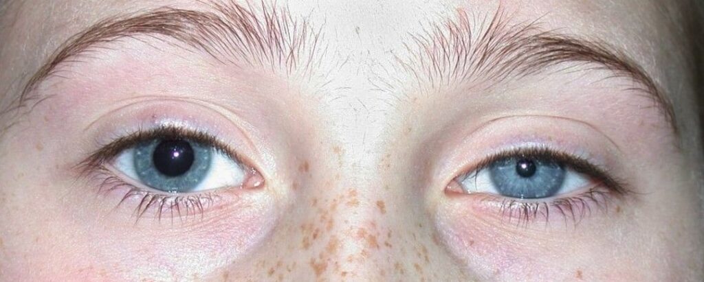
In addition, look for mild ptosis, an elevated lower lid and apparent enophthalmos. Physiologic anisocoria can mimic some of these findings, therefore pharmacologic testing helps.
All-fit Slit-Lamp Adapter
Record and share exactly what you see at the slit lamp. One adapter fits any slit lamp or surgical microscope — and any smartphone.
From Choroida — the team behind this siteApraclonidine Testing Before Horner Localization
Apraclonidine 0.5% has weak alpha-1 activity that acts on denervated, upregulated receptors. As a result, the Horner pupil dilates and the lid often rises, reversing the anisocoria. Cocaine 4% to 10% is the older test, and it fails to dilate the Horner pupil.
Kardon and colleagues found that post-cocaine anisocoria of 0.8 mm or more strongly supported Horner syndrome. However, apraclonidine may give a false negative soon after onset, because receptor upregulation takes time.
Hydroxyamphetamine and Horner Localization
Hydroxyamphetamine 1% releases stored norepinephrine from intact third-order nerve terminals. Therefore, a pupil that dilates poorly suggests a third-order (postganglionic) lesion. Normal dilation suggests a first- or second-order lesion, although the test cannot separate those two.
Maloney and colleagues reported good localizing accuracy for this approach, and clinicians should wait at least 48 to 72 hours after cocaine or apraclonidine before testing. Also, the drug is hard to obtain in many countries, which limits its routine use.
Anhidrosis Pattern in Horner Localization
Sweating clues help when present, although patients rarely notice them. A first-order lesion may reduce sweating over the whole ipsilateral body, while second-order lesions typically affect the face and neck on that side. In contrast, third-order lesions spare most facial sweating, because those fibers follow the external carotid artery.
First-Order Clues
Look carefully for brainstem and cord signs. Lateral medullary infarction may add vertigo, ataxia, dysphagia and crossed sensory loss. Next, cervical cord lesions such as syringomyelia can produce limb weakness or dissociated sensory loss.
Second-Order Clues
Ask about smoking, cough, shoulder pain, thyroid disease and previous neck or chest surgery. For example, an apical lung tumor can cause arm pain and hand weakness with the Horner syndrome. Central lines, chest tubes and neck trauma also injure this segment.
Third-Order Clues
Ipsilateral neck, face or head pain points toward internal carotid dissection. In addition, cavernous sinus disease may add third, fourth or sixth nerve palsy and facial numbness (see third nerve palsy). Cluster headache can also leave a postganglionic Horner syndrome after attacks.
Pain and Acute Onset in Horner Localization
A new painful Horner syndrome suggests carotid dissection until imaging excludes it. Dissection carries a risk of ischemic stroke, therefore same-day vascular imaging is appropriate. Do not delay imaging while waiting for pharmacologic tests.
Imaging by Order
For first-order lesions, request MRI of the brain and cervical spine. Second-order lesions need imaging of the lung apex and neck, usually with CT or MRI. Third-order lesions need CT or MR angiography of the head and neck, with attention to the cavernous sinus.
When the order remains uncertain, image the whole pathway from hypothalamus to upper chest. Davagnanam and colleagues proposed a combined clinical, pharmacologic and imaging algorithm for this situation.
Children and Horner Localization
Congenital cases may show a lighter iris on the affected side. However, acquired Horner syndrome in a child requires a search for neuroblastoma. Mahoney and colleagues supported imaging of the head, neck and chest, plus urine catecholamine testing.
Counseling the Patient
Explain that the droop and small pupil rarely affect vision. Then explain why imaging matters, because the cause carries the real risk. Finally, advise urgent return for new headache, weakness, speech change or visual loss.
Pitfalls to Avoid
- Do not rely on hydroxyamphetamine alone to exclude a central lesion
- Never repeat drop testing within 72 hours of another test
- Do not attribute a new painful Horner syndrome to migraine
- Also exclude a pharmacologically dilated fellow pupil (see pharmacologic mydriasis)


Document what you see
Two smartphone imaging tools built for everyday clinic use — one for the slit lamp, one for the fundus.
From Choroida — the team behind this siteReferences
- Kardon RH, Denison CE, Brown CK, Thompson HS. Critical evaluation of the cocaine test in the diagnosis of Horner’s syndrome. Arch Ophthalmol. 1990;108:384-387.
- Koc F, Kavuncu S, Kansu T, et al. The sensitivity and specificity of 0.5% apraclonidine in the diagnosis of oculosympathetic paresis. Br J Ophthalmol. 2005;89:1442-1444.
- Maloney WF, Younge BR, Moyer NJ. Evaluation of the causes and accuracy of pharmacologic localization in Horner’s syndrome. Am J Ophthalmol. 1980;90:394-402.
- Davagnanam I, Fraser CL, Miszkiel K, Daniel CS, Plant GT. Adult Horner’s syndrome: a combined clinical, pharmacological, and imaging algorithm. Eye. 2013;27:291-298.
- Mahoney NR, Liu GT, Menacker SJ, et al. Pediatric Horner syndrome: etiologies and roles of imaging and urine studies to detect neuroblastoma and other responsible mass lesions. Am J Ophthalmol. 2006;142:651-659.
Test yourself
A few questions straight from this article.
-
In Horner syndrome, the first-order sympathetic neuron runs from the hypothalamus to the ciliospinal center at which spinal level?
The first-order neuron runs from the hypothalamus through the brainstem to the ciliospinal center at C8 to T2. -
Which structure does the second-order sympathetic neuron cross before reaching the superior cervical ganglion in the Horner pathway?
The second-order neuron leaves the cord, crosses the lung apex and ascends to the superior cervical ganglion, which is why apical tumors and chest procedures injure it. -
How does the anisocoria of Horner syndrome change in dim light?
The Horner pupil dilates slowly in darkness, so anisocoria increases in dim light, and dilation lag in the first seconds of darkness also supports the diagnosis. -
What happens to a Horner pupil after instillation of apraclonidine 0.5%?
Apraclonidine's weak alpha-1 activity acts on denervated, upregulated receptors, so the Horner pupil dilates and the lid often rises, reversing the anisocoria. -
Why may apraclonidine testing give a false negative soon after the onset of Horner syndrome?
Apraclonidine relies on denervation supersensitivity, and receptor upregulation takes time, so testing soon after onset may be falsely negative. -
In Horner syndrome, a pupil that dilates poorly after hydroxyamphetamine 1% suggests a lesion of which neuron?
Hydroxyamphetamine releases stored norepinephrine from intact third-order terminals, so poor dilation suggests a postganglionic lesion; normal dilation cannot separate first- from second-order lesions. -
A Horner syndrome with vertigo, ataxia, dysphagia and crossed sensory loss localizes to which neuron order?
Lateral medullary infarction may add vertigo, ataxia, dysphagia and crossed sensory loss, which are first-order (central) clues. -
A patient presents with a new painful Horner syndrome. What is the appropriate next step?
A new painful Horner syndrome suggests carotid dissection, which carries a risk of ischemic stroke, so same-day vascular imaging should not wait for drop tests. -
Which imaging is requested for a first-order Horner syndrome?
First-order lesions need MRI of the brain and cervical spine; second-order lesions need lung apex and neck imaging, and third-order lesions need CT or MR angiography. -
An acquired Horner syndrome in a child requires a search for which tumor?
Acquired Horner syndrome in a child requires a neuroblastoma search with head, neck and chest imaging plus urine catecholamine testing.

Writes and reviews the clinical articles on Educate. Every post is checked against peer-reviewed sources before it goes live.