Focal and grid laser was the standard treatment for diabetic macular edema for three decades before anti-VEGF drugs arrived.
It still has a role in eyes with noncenter-involving edema, in selected patients who cannot receive injections, and as an adjunct.
Knowing the technique helps in choosing patients and avoiding complications.
How Focal and Grid Laser Works
Laser burns the retinal pigment epithelium and outer retina, and the resulting scar reduces edema over months.
Possible mechanisms include reduced oxygen demand, improved oxygen supply to the inner retina, and release of RPE factors that restore the barrier.
The exact mechanism remains uncertain, though the effect is clear in trials (see diabetic macular edema).
Focal Versus Grid Treatment
Focal treatment targets individual leaking microaneurysms, usually found by fluorescein angiography.
Grid treatment places a pattern of spots across areas of diffuse leakage or nonperfusion in the macula.
Many eyes need a combination, with focal spots to leaking points and a grid to diffuse edema (see fluorescein angiography patterns).
Fundus Explorer Pro
Photograph the retinal findings described here with the phone already in your pocket — 22 D optics and built-in illumination in one handheld unit.
From Choroida — the team behind this siteEvidence for Focal and Grid Laser
The ETDRS showed that focal laser reduced the risk of moderate visual loss by about half in eyes with clinically significant macular edema.
After this trial, laser became the standard care for years.
Later DRCR Network trials found that ranibizumab with prompt or deferred laser gave better vision than laser alone for center-involving edema.
Therefore, anti-VEGF drugs now lead, with laser as an adjunct.
Technique
Use a contact lens and a small spot size, 50 to 100 micrometers, with short duration, 0.05 to 0.1 second.
Aim for a mild burn that is barely visible, and avoid confluent burns.
Treat leaking microaneurysms between 500 and 3000 micrometers from the foveal center.
For grid treatment, space spots one burn width apart, and keep them outside the foveal avascular zone.
Complications of Focal and Grid Laser
- Enlarging laser scars that reach the fovea over years
- Paracentral scotomas and loss of contrast
- Choroidal neovascularization from a break in Bruch membrane
- Transient increase in edema in the first weeks
Careful settings and wide spacing lower these risks.
Current Role of Focal and Grid Laser
Use laser for noncenter-involving clinically significant edema, especially when vision is good.
Also use it when patients cannot attend frequent injection visits or when cost is a barrier.
In combination with injections, laser may reduce the number of injections in some eyes (see diabetic retinopathy management).
An Example Case
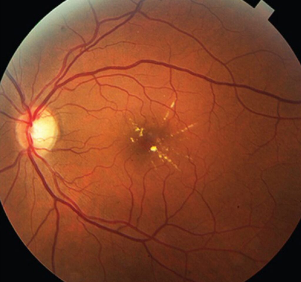
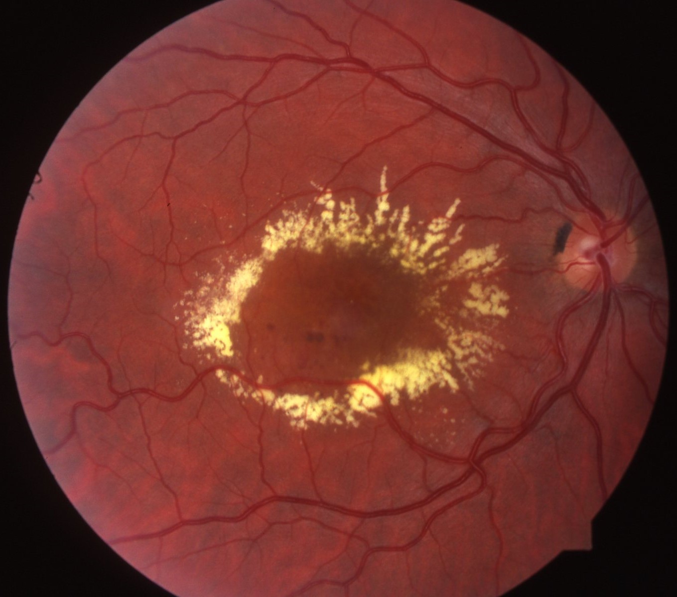
A 58-year-old woman has type 2 diabetes and noncenter-involving edema with hard exudates in a circinate ring.
Her acuity is 20/25, and OCT shows thickening 800 micrometers temporal to the fovea.
Fluorescein angiography shows leaking microaneurysms at the center of the ring.
She receives focal laser to the leaking points, and the edema and exudates fade over three months.
Her vision stays at 20/25, and she avoids injections.
Questions Patients Ask
Patients ask whether the laser will improve their vision, and the answer is that it aims to stop vision from getting worse.
They also ask whether it hurts, and many feel a brief stinging sensation.
Because the effect takes weeks to months, set realistic expectations.
Pitfalls to Avoid
- Treating microaneurysms within 500 micrometers of the foveal center
- Using large or intense burns that enlarge into scars
- Applying laser in eyes with center-involving edema without considering injections
- Overlooking the need for good glycemic and blood pressure control
Looking Ahead
Subthreshold laser techniques may reduce scarring and keep some of the benefit.
Combination approaches will continue to evolve as new drugs appear.
However, laser stays a low-cost option in settings with limited access to injections.
Practical Points
Obtain fluorescein angiography and OCT before treatment, so that targets are clear.
Then record the spot size, power, and number of burns in the chart.
Also schedule review at three months, since retreatment may be needed.
Preparing the Patient
Check blood pressure, glycemic control, and kidney function before treatment, since these factors influence edema.
Then explain that laser needs several visits and may not improve vision.
Also warn the patient about possible scotomas and slow resolution.
Follow-Up After Laser
Review at three to four months with OCT.
If edema persists, retreat only microaneurysms outside the safe zone.
Because injections now lead for center-involving edema, consider switching to an anti-VEGF agent when thickness rises.


Document what you see
Two smartphone imaging tools built for everyday clinic use — one for the slit lamp, one for the fundus.
From Choroida — the team behind this siteReferences
- Early Treatment Diabetic Retinopathy Study Research Group. Photocoagulation for diabetic macular edema. Early Treatment Diabetic Retinopathy Study report number 1. Arch Ophthalmol. 1985;103:1796-1806.
- Elman MJ, Aiello LP, Beck RW, et al. Randomized trial evaluating ranibizumab plus prompt or deferred laser or triamcinolone plus prompt laser for diabetic macular edema. Ophthalmology. 2010;117:1064-1077.
- Flaxel CJ, Adelman RA, Bailey ST, et al. Diabetic retinopathy preferred practice pattern. Ophthalmology. 2020;127:P66-P145.
- American Academy of Ophthalmology. Basic and Clinical Science Course, Section 12: Retina and Vitreous. AAO; current edition.

Writes and reviews the clinical articles on Educate. Every post is checked against peer-reviewed sources before it goes live.