Keratoconus in children behaves differently from the adult disease, and the differences affect how soon doctors should offer treatment.
It is more advanced at diagnosis, it progresses faster, and it is more likely to need a graft if doctors leave it untreated.
Early detection is also the best tool for protecting vision in a growing eye.
Why Is Keratoconus in Children More Aggressive?
- Collagen in young corneas is less stiff, and the cone can also steepen quickly during puberty
- Allergic eye disease and eye rubbing are common in children and drive the condition (see vernal keratoconjunctivitis)
- Associations with Down syndrome, Leber congenital amaurosis, and connective tissue disease are more frequent in the young
- Family history and genetic factors act earlier in some patients
Studies show that doctors often diagnose children at a more advanced stage than adults, since symptoms are overlooked.
Keratoconus in Children: How Children Present
- Frequent changes in glasses, and a rising astigmatism
- Poor vision that does not correct well, and an unexplained drop in school performance
- Squinting, eye rubbing, and sensitivity to light
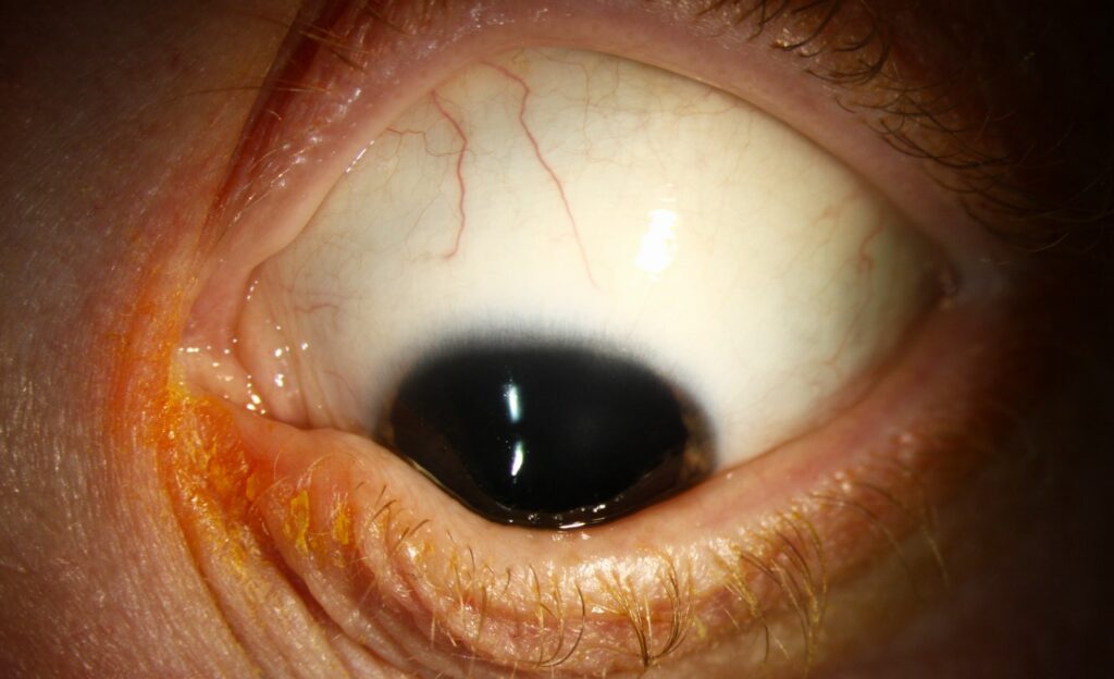
- Acute hydrops in advanced disease (see acute corneal hydrops)
Young children may not complain, and a school screening or a routine refraction may find the problem.
All-fit Slit-Lamp Adapter
Record and share exactly what you see at the slit lamp. One adapter fits any slit lamp or surgical microscope — and any smartphone.
From Choroida — the team behind this siteDetection
- Retinoscopy and keratometry show irregular astigmatism and steep readings
- Corneal topography and tomography reveal the cone, and are the main tools for diagnosis and follow-up (see corneal topography)
- Slit lamp signs, such as Vogt striae, may be absent in early disease
- Also keep a high index of suspicion in children with allergic disease, Down syndrome, or a family history
Clinicians advise screening of siblings and of children with risk factors.
Keratoconus in Children: Treatment Principles
Because progression in children is fast, many specialists advise cross-linking as soon as progression is documented, and some at the time of diagnosis in young patients.
- Cross-linking: strengthens the cornea and arrests progression in most cases; the KERALINK trial in patients aged 10 to 16 showed that it halted progression compared with standard care (see the references and corneal cross-linking)
- Spectacles and soft lenses in early disease
- Rigid gas permeable and scleral lenses for visual rehabilitation (see scleral lens fitting)
- Control of allergy and rubbing, which is essential
- Keratoplasty in advanced disease, with a higher rate of graft rejection in children
Cross-linking does not cure the condition, but it prevents a worse outcome.
Special Challenges
- Children may also cooperate poorly with topography and with the procedure, and some need sedation or general anesthesia
- Epithelium-on and accelerated protocols are attractive for children, though researchers are still building the evidence (see epi-on cross-linking)
- Follow-up every three to six months is needed, since progression can occur at any time until the late twenties or thirties
Counselling Families
Explain the disease in simple terms, and stress that rubbing the eyes worsens it.
In addition, describe the benefits of treatment, and the need for regular follow-up.
Refer to a center with experience in pediatric cross-linking when it is not available locally.
Why Early Treatment Pays Off
Waiting for a child to reach the stage where vision is poor means accepting a higher chance of scarring, a thinner cornea, and a possible graft.
Cross-linking at an earlier stage preserves corneal tissue and offers more options for vision correction later.
Studies suggest that the rate of progression in the first years after diagnosis is high in young patients, and that treatment in this period gives the best chance to halt it.
Keratoconus in Children: Follow-Up Plan After Treatment
- Review at one week, one month, three months, and six months, with topography at each stage after the first month
- Continue annual review into adulthood, since new progression can occur
- Update the spectacle or lens prescription as the cornea settles over several months
- Reinforce the advice on eye rubbing and allergy treatment at every visit
Tips for the Clinic
- Ask about rubbing, atopy, sleep position, and family history in every child with changing astigmatism
- Obtain a baseline topography even in children who are not yet symptomatic when there is a strong risk
- Involve the pediatrician when you suspect a systemic association
Families who understand the reason for treatment are more likely to cooperate with follow-up.
Parents are often the first to notice squinting or frequent rubbing, so ask them directly during the history, and ask about the child’s sleeping position and use of screens.
For the latest studies, see the PubMed results for keratoconus in children.


Document what you see
Two smartphone imaging tools built for everyday clinic use — one for the slit lamp, one for the fundus.
From Choroida — the team behind this siteReferences
- Larkin DFP, Chowdhury K, Burr JM, et al. Effect of corneal cross-linking vs standard care on keratoconus progression in young patients: the KERALINK randomized controlled trial. Ophthalmology. 2021;128:1516-1526.
- Chatzis N, Hafezi F. Progression of keratoconus and efficacy of pediatric corneal collagen cross-linking in children and adolescents. J Refract Surg. 2012;28:753-758.
- Mukhtar S, Ambati BK. Pediatric keratoconus: a review of the literature. Int Ophthalmol. 2018;38:2257-2266.
- American Academy of Ophthalmology. Basic and Clinical Science Course, Section 8: External Disease and Cornea. AAO; current edition.

Writes and reviews the clinical articles on Educate. Every post is checked against peer-reviewed sources before it goes live.