AI in retinal OCT interpretation is already part of the workflow in many clinics, from automatic layer segmentation to fluid detection.
Deep learning models can also read scans quickly and consistently, and they may help busy services to find patients who need treatment.
They do not replace the clinician, because they fail in ways that need to be understood.
What AI in Retinal OCT Interpretation Does
- Segmentation: finding retinal layers and measuring thickness
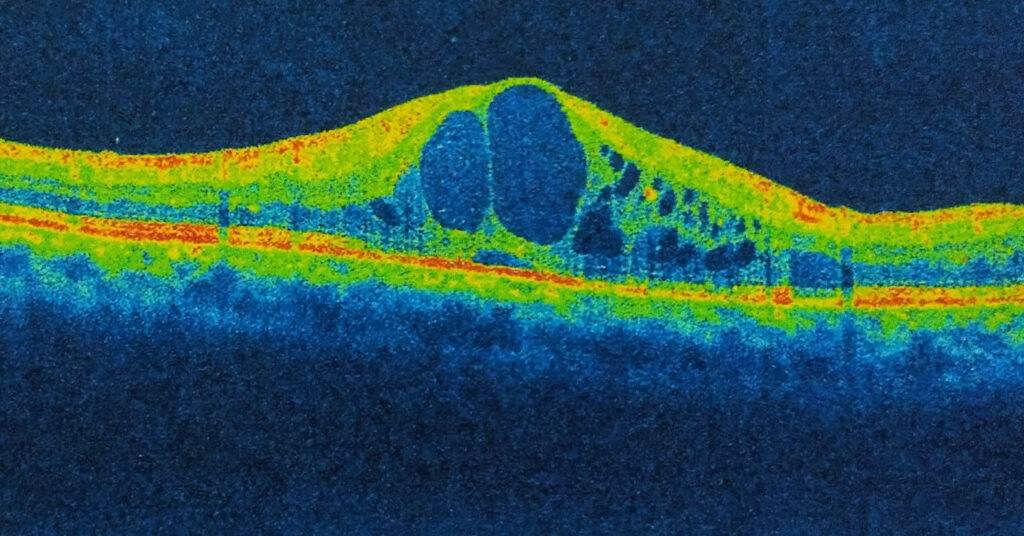
- Fluid quantification: outlining intraretinal fluid, subretinal fluid, and pigment epithelial detachment
- Atrophy measurement: mapping the area of geographic atrophy and outer retinal loss
- Classification: assigning a scan to a disease category or a referral urgency
- Prediction: estimating the response to treatment or the risk of progression
AI in Retinal OCT Interpretation: Landmark Studies
A study of the Moorfields service showed that a deep learning system could recommend referral decisions for more than 50 retinal conditions on OCT, with accuracy matching expert clinicians (see the references).
Another large study trained models on tens of thousands of scans and identified treatable diseases, such as choroidal neovascularization and diabetic edema (see the references).
Also, developers can adapt foundation models trained on huge numbers of retinal images to new tasks with little data (see the references).
Fundus Explorer Pro
Photograph the retinal findings described here with the phone already in your pocket — 22 D optics and built-in illumination in one handheld unit.
From Choroida — the team behind this siteHow AI Helps in the Clinic
- Triage of urgent cases, such as new neovascular AMD, within busy services
- Support for anti-VEGF decisions through objective fluid volumes (see oct angiography)
- Quality checks of scan acquisition
- Teaching tools for trainees
- Population screening for diabetic edema and glaucoma in primary care
AI in Retinal OCT Interpretation: Problems and Limits
- Dataset bias: models trained on one device or population may also fail on another
- Image quality: poor scans, media opacities, and artifacts lead to errors
- Rare diseases: few examples make models unreliable for unusual cases
- Black box: many models do not explain their reasoning, which makes errors hard to detect
- Automation bias: clinicians may also over-trust a confident output
Validation and Regulation
Clinicians test good tools on external datasets, in prospective trials, and in real use.
Also, regulators in several regions have cleared AI systems for diabetic screening, and approval for OCT tools is growing.
A model that works well in a paper may not work in a clinic, so audit locally.
AI in Retinal OCT Interpretation: Practical Guidance
- Use AI output as a second reader and not as a final answer
- Check the segmentation lines and fluid maps yourself on critical scans
- Know the device and software version, and the population on which developers validated it
- Keep a record of disagreements, so you can track errors
Ethical Points
Protect patient data, and consent should cover its use in training.
In addition, test algorithms for fairness across ages, ethnic groups, and devices.
AI in Retinal OCT Interpretation: What Comes Next
Multimodal models that combine OCT, fundus photos, and clinical data may predict disease and response to treatment, and may guide personalized treatment plans (see AI for diabetic retinopathy screening).
Example Workflow
Staff acquire a scan in a primary care or optometry setting, and software flags fluid at the fovea with a high confidence.
The system also refers the patient for same-week review, where a clinician confirms the finding and starts treatment.
Without the tool, the scan might have waited in a queue for weeks, so the main value is speed and consistency.
Measuring Fluid Objectively
Fluid volumes from AI segmentation allow the clinician to compare scans over time, and treatment decisions can be based on trends, and not on a single thickness number.
Models can also separate fluid in the retina from fluid under it, which may relate to different outcomes.
Data Privacy and Governance
Hospitals must secure patient data, set clear rules for who can review results, and decide how to validate algorithm updates.
Vendors should also disclose the data used for training, and the performance in subgroups.
Continuing Education
Clinicians should understand the basics of how models are trained and tested, and include short sessions in training programs, so that they can use the tools wisely and teach patients to trust them within limits.
Local audit of performance, with simple monthly reports of sensitivity and false alarms, shows whether a tool is helping or adding work.
Common Pitfalls in Daily Use
- Automatic thickness maps can be wrong in eyes with schisis, high myopia, or large detachments, so check the segmentation lines
- The software may flag a scan with low signal as normal by mistake
- Differences between devices change the numbers, so compare scans from the same machine
- Fluid volumes depend on the software version, so do not mix versions in a follow-up series
Teaching staff to look for these pitfalls prevents most errors and keeps the benefit of the tool.


Document what you see
Two smartphone imaging tools built for everyday clinic use — one for the slit lamp, one for the fundus.
From Choroida — the team behind this siteReferences
- De Fauw J, Ledsam JR, Romera-Paredes B, et al. Clinically applicable deep learning for diagnosis and referral in retinal disease. Nat Med. 2018;24:1342-1350.
- Kermany DS, Goldbaum M, Cai W, et al. Identifying medical diagnoses and treatable diseases by image-based deep learning. Cell. 2018;172:1122-1131.
- Zhou Y, Chia MA, Wagner SK, et al. A foundation model for generalizable disease detection from retinal images. Nature. 2023;622:156-163.
- American Academy of Ophthalmology. Basic and Clinical Science Course, Section 12: Retina and Vitreous. AAO; current edition.

Writes and reviews the clinical articles on Educate. Every post is checked against peer-reviewed sources before it goes live.