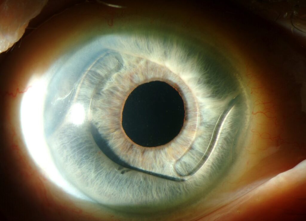
An anterior chamber intraocular lens sits in front of the iris, in the angle of the eye, and is used when the capsular bag cannot support a lens.
Early rigid designs caused serious problems, but modern flexible open-loop lenses have a good safety record in suitable eyes.
It remains a useful, quick option, though other techniques compete with it.
When Is an Anterior Chamber Intraocular Lens Used?
- Aphakia after complicated cataract surgery with loss of capsular support
- Dislocated or subluxated lens or IOL (see dislocated intraocular lens)
- Secondary implantation in aphakic eyes, after intracapsular surgery or trauma
- As a primary option in cases where a posterior chamber lens cannot be placed, and where other techniques are not available
In older patients with a healthy cornea and angle, it gives a quick solution with less surgery time.
Lens Designs
- Closed-loop lenses, an early design that caused problems, and are also no longer used
- Flexible open-loop lenses, with four-point fixation in the angle, which are also the standard today
- Iris-claw lenses, fixed to the iris, which are not placed in the angle, and have a different profile
- Angle-supported lenses that come in sizes suited to the eye
Sizing is important, so the lens is typically chosen at 1 millimeter more than the white-to-white distance.
All-fit Slit-Lamp Adapter
Record and share exactly what you see at the slit lamp. One adapter fits any slit lamp or surgical microscope — and any smartphone.
From Choroida — the team behind this siteSurgical Technique
- First, construct a clear corneal or scleral tunnel incision of adequate size
- Fill the chamber with viscoelastic, and perform an anterior vitrectomy if there is vitreous
- Constrict the pupil with miotic drops, and insert the lens carefully into the angle
- Make a peripheral iridotomy to prevent pupillary block
- Check the position, and that the haptics lie flat without tucking the iris
- Remove the viscoelastic, and then close the wound
Results of Anterior Chamber Intraocular Lens
Studies comparing ACIOLs with sutured posterior chamber lenses in eyes with poor capsular support show similar visual outcomes and complication rates in modern series (see the references).
Also, the American Academy of Ophthalmology review concluded that open-loop ACIOLs and sutured PCIOLs are both acceptable options (see the references).
Complications of Anterior Chamber Intraocular Lens
- Corneal endothelial loss and bullous keratopathy, more frequent with poor sizing or contact (see pseudophakic bullous keratopathy)
- Glaucoma and angle damage, from chafing or synechiae
- Uveitis-glaucoma-hyphema syndrome (see uveitis-glaucoma-hyphema syndrome)
- Cystoid macular edema, from inflammation and vitreous traction
- Pupil ovalization and iris tuck
- Incorrect size, with lens rotation or too much movement
Follow-up of the endothelium and pressure is also needed.
Contraindications of Anterior Chamber Intraocular Lens
- Poor corneal endothelium or low cell count
- Narrow or damaged angle, or glaucoma
- Chronic uveitis
- Shallow anterior chamber
- Eyes at risk of rubeosis
Alternatives
- Scleral-fixated lenses, with sutures or glue (see scleral-fixated IOL)
- Intrascleral haptic fixation, such as the Yamane technique
- Iris-fixated lenses
- Contact lens or glasses
The choice depends on the surgeon’s skill, the eye, and the patient.
Counselling
Explain the reasons for the lens, the expected vision, and the need for lifelong follow-up, including pressure and corneal checks.
Sizing Pearls
- Measure the white-to-white distance with calipers or a device, and add about one millimeter for lens length
- A lens that is too small can also rotate and cause chafing, and one that is too large causes pressure on the angle and ovalization of the pupil
- Check the final position with gonioscopy or anterior segment imaging, when available (see ultrasound biomicroscopy)
Case Example
A 74-year-old man has a posterior capsule rupture with loss of the nucleus into the vitreous, treated by vitrectomy.
Two months later, with a clear cornea and a healthy angle, the surgeon implants an open-loop anterior chamber lens with an iridotomy.
Vision reaches 20/30, and the team follows the patient with endothelial counts and pressure checks.
Follow-Up for Anterior Chamber Intraocular Lens
- Review at one day, one week, and one month, then every six months
- Measure pressure and examine the angle for synechiae
- Perform specular microscopy at yearly intervals when possible
- Treat inflammation and edema early, and then consider lens exchange for chafing or corneal decompensation (see IOL exchange)
Who Does Well?
Older patients with a healthy cornea and a wide angle usually do well, and younger patients with long life expectancy may be better served by lenses fixed to the sclera or iris, because of the long exposure of the corneal endothelium.
Always document the lens model, size, and the haptic position in the record, so that later surgeons can plan lens exchange or removal if complications arise.
Remind patients that sudden pain, redness, or reduced vision after surgery needs prompt review, since corneal and pressure problems can develop quietly and are easier to manage early.


Document what you see
Two smartphone imaging tools built for everyday clinic use — one for the slit lamp, one for the fundus.
From Choroida — the team behind this siteReferences
- Wagoner MD, Cox TA, Ariyasu RG, Jacobs DS, Karp CL. Intraocular lens implantation in the absence of capsular support: a report by the American Academy of Ophthalmology. Ophthalmology. 2003;110:840-859.
- Donaldson KE, Gorscak JJ, Budenz DL, et al. Anterior chamber and sutured posterior chamber intraocular lenses in eyes with poor capsular support. J Cataract Refract Surg. 2005;31:903-909.
- Kohnen T, Koch DD, eds. Cataract and Refractive Surgery. Springer; 2005.
- American Academy of Ophthalmology. Basic and Clinical Science Course, Section 11: Lens and Cataract. AAO; current edition.

Writes and reviews the clinical articles on Educate. Every post is checked against peer-reviewed sources before it goes live.