
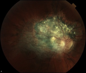
Some fundus lesions look far more sinister than they are.
A grey, slightly raised patch near the optic disc, dragging the retinal vessels into a corkscrew, can send a child straight down a work-up for melanoma or retinoblastoma.
Yet the lesion is benign, congenital, and will never spread.
Combined Hamartoma of the Retina and RPE (CHRRPE) is a rare, usually congenital, benign tumour that disorganizes the full thickness of the retina and retinal pigment epithelium.
It is frequently mistaken for a sinister pigmented lesion — a choroidal melanoma or retinoblastoma — or, at the other extreme, dismissed as an “idiopathic epiretinal membrane.”
Most cases are unilateral and sporadic. A minority, however, signal neurofibromatosis type 2 (NF2).
It is also a quiet cause of unexplained amblyopia, strabismus, and painless visual loss in a child with an otherwise normal anterior segment.
Benign yet visually disabling — and occasionally the first clue to NF2 — CHRRPE rewards the clinician who can recognise its fundus and OCT signature.
What Is Combined Hamartoma of the Retina and RPE?
CHRRPE is a benign, disorganized proliferation of retinal glial tissue, blood vessels, RPE, and a contractile epiretinal membrane. The term was coined by Gass in 1973.
It combines three tissues, and each leaves a fingerprint on the fundus:
- RPE — variable pigmentation, grey to charcoal.
- Retinal vasculature — tortuous, dragged vessels.
- Glial and membranous tissue — an overlying grey-white veil.
The classic lesion is slightly elevated and ill-defined, blending imperceptibly with the surrounding retina. A grey-white glial veil sits over the pigment.
Just as telling is what is absent: no choroidal or RPE atrophy at the margin, no subretinal fluid, no haemorrhage, and no vitritis.
Three locations are described, in order of frequency:
- Juxtapapillary or peripapillary — the most common.
- Macular.
- Peripheral.
A grey, ill-defined, full-thickness lesion that fades into normal retina — rather than a discrete choroidal mass — is the first clue that you are looking at a hamartoma, not a melanoma.
Epidemiology and Associations
CHRRPE is rare, and its true incidence is unknown.
It usually presents in childhood or young adulthood, with no clear sex predilection, and is almost always unilateral.
In a large case series, the presenting picture broke down roughly as follows:
- Decreased vision — about 40%.
- Strabismus — about 28%.
- Incidental discovery — about 23%.
Macular and extramacular locations were roughly equal in that series.
Most cases are sporadic, but several systemic associations are worth naming:
- Neurofibromatosis type 2 (NF2) — the strongest association.
- Neurofibromatosis type 1 (NF1) — weaker.
- Gorlin (basal-cell nevus) syndrome.
- Juvenile nasopharyngeal angiofibroma.
- Incontinentia pigmenti.
- Branch retinal vein occlusion.
- X-linked juvenile retinoschisis.
- Optic disc pit and coloboma.
Bilateral or multifocal CHRRPE is particularly important — it should prompt a work-up for NF2.
Fundus Explorer Pro
Photograph the retinal findings described here with the phone already in your pocket — 22 D optics and built-in illumination in one handheld unit.
From Choroida — the team behind this sitePathogenesis
CHRRPE is best understood as a congenital hamartomatous overgrowth of retina and RPE, with a contractile glial epiretinal membrane as the central driver.
Modern OCT reappraisal has refined that picture. The lesion behaves as a thickened retinal mass secondary to focal vitreoretinal traction, with a primary origin in the inner retinal layers and progressive extension outward.
Contraction of the membrane then sets off the visible sequence:
- Retinal folding.
- Vascular tortuosity and dragging.
- Macular distortion.
That last step — traction-driven macular distortion — is the main mechanism of visual loss.
Clinical Presentation
Symptoms depend heavily on lesion location and the patient’s age.
Adults and older children may report:
- Painless decreased vision.
- Metamorphopsia.
- Strabismus.
- Leukocoria — rare.
Many lesions are entirely asymptomatic and found incidentally.
In young children, the presentation is often indirect: amblyopia or strabismus in an eye that looks quiet on external and anterior-segment examination.
A child with unexplained amblyopia and a normal front of the eye deserves a careful dilated fundus examination before the diagnosis is left as “idiopathic.”
Examination Findings
On dilated fundoscopy, the lesion is characteristic once you know the pattern.
The key features include:
- A slightly elevated, grey-to-charcoal, ill-defined lesion that blends into normal retina.
- Retinal vascular tortuosity and dragging.
- Corkscrew intralesional vessels.
- An overlying epiretinal membrane or gliosis exerting traction.
The negatives are equally diagnostic. Characteristically absent are:
- Subretinal fluid.
- Haemorrhage.
- Choroidal involvement.
- Vitritis.
A grey lesion dragging the vessels into a corkscrew, with no subretinal fluid and a quiet choroid, is the fundus signature of CHRRPE.
Why It Is So Often Misdiagnosed
CHRRPE sits in a diagnostic blind spot, and it gets missed in two opposite directions.
In adults, it is under-called — overlooked as an “idiopathic epiretinal membrane.” The giveaway that separates it from a simple ERM is the underlying pigment and the deep vascular disorganization.
In children, it is over-called — mistaken for a choroidal melanoma or retinoblastoma, triggering unnecessary alarm and work-up.
The OCT traction signs help, but a note of caution belongs here:
- The inner-retinal “peaks” and the “omega” configuration are characteristic — but they are not pathognomonic.
- No single sign confirms the diagnosis in isolation.
The safest approach is to read the whole picture — fundus appearance, OCT, and angiography together — rather than lean on any one finding.
Diagnostic Evaluation
Fundus Photography
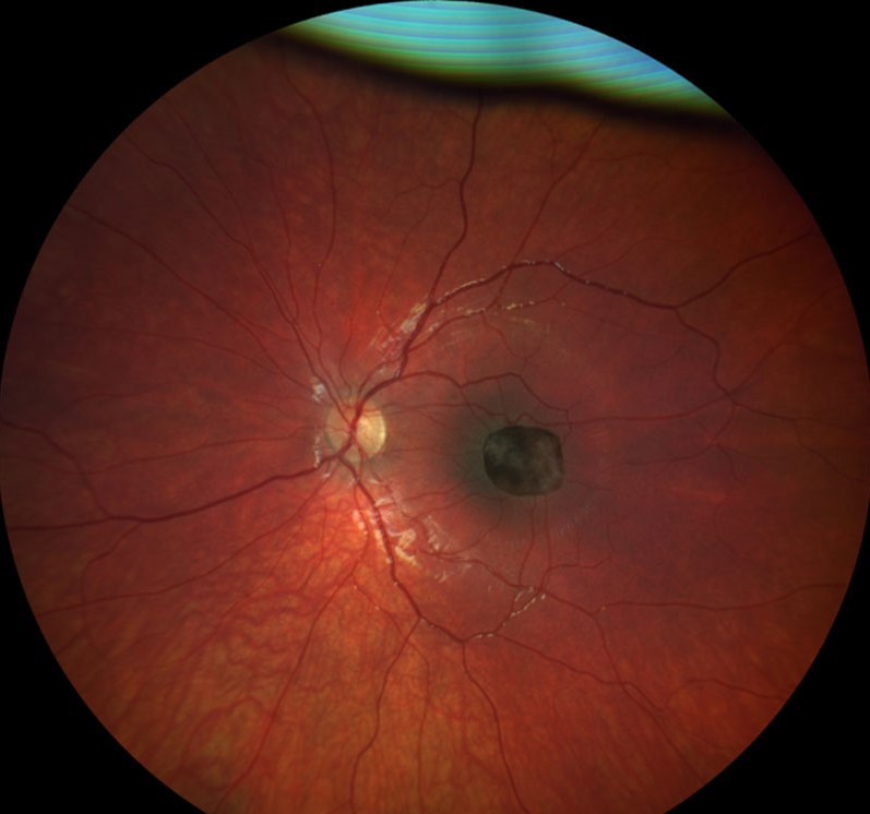
Colour fundus photography documents the grey glial veil, the variable pigment, and the dragged, tortuous vessels — and provides the baseline for tracking change over time.
Optical Coherence Tomography
OCT is the key confirmatory investigation.
Typical findings include:
- Inner-retinal “mini-peaks” — a sawtooth contour from superficial traction.
- Full-thickness “maxi-peaks” — folds driven by vitreoretinal traction.
- Retinal thickening and full-thickness disorganization.
- Posterior optical shadowing that obscures the outer retinal layers.
A family of descriptive signs has grown up around these appearances — shark-tooth, omega, and double-retina among them.
The full-thickness peaks and disorganization are the finding that most reliably separates CHRRPE from a simple epiretinal membrane.
Fluorescein Angiography
FA highlights the abnormal vasculature:
- Tortuous, corkscrew intralesional vessels.
- An early fine capillary network.
- Late leakage and staining.
OCT Angiography and B-Scan
OCT-A is an emerging adjunct that can show the abnormal, tortuous intralesional vasculature without dye.
When the media are hazy, B-scan ultrasonography demonstrates a mildly elevated, non-calcified lesion — a useful point of separation from calcified retinoblastoma.
Differential Diagnosis
Conditions that may mimic CHRRPE include:
- Idiopathic epiretinal membrane — lacks the underlying pigment, the deep vascular disorganization, and the full-thickness OCT peaks.
- Choroidal melanoma — choroidal in origin, often with subretinal fluid; CHRRPE spares the choroid and shows no SRF.
- Retinoblastoma — chalky white and calcified; CHRRPE is non-calcified, grey, and quiet.
- CHRPE — flat, well-demarcated, jet-black with lacunae; CHRRPE is elevated, ill-defined, and traction-related.
- Myelinated nerve fibres — feathered white along the nerve fibre layer, with no traction or pigment.
- Optic disc anomaly or coloboma.
- X-linked juvenile retinoschisis — schisis cavities on OCT.
Distinguishing CHRRPE from melanoma and retinoblastoma spares the patient unnecessary alarm — and sometimes an unnecessary enucleation.
Management
Observation
Observation is first-line for most patients.
It is appropriate for lesions that are:
- Extramacular.
- Asymptomatic.
- Stable.
In children, observation is paired with active amblyopia therapy and strabismus management, alongside serial OCT and photography to detect progression.
Surgery
Pars plana vitrectomy with membrane peel is reserved for progressive macular traction and distortion with declining vision.
Counsel patients realistically: visual gains are limited and unpredictable, because the retina beneath the membrane is intrinsically disorganized. Relieving traction does not restore normal architecture.
Systemic Evaluation
Systemic associations should be evaluated and monitored, with formal NF2 evaluation where the clinical picture — particularly bilateral or multifocal disease — indicates it.
For most patients, careful observation with amblyopia care does more good than early surgery.
Prognosis
CHRRPE is benign. It does not metastasise and does not undergo malignant transformation.
Visual outcome is driven almost entirely by location, and macular lesions do worse. In a large case series:
- Presenting visual acuity was 20/200 or worse in about 47% of eyes.
- Poor vision occurred in roughly 69% of macular versus about 25% of extramacular tumours.
- Continued visual decline was more frequent in macular (about 60%) than in extramacular (about 13%) lesions.
The two main threats to vision are amblyopia in children and progressive epiretinal membrane traction — both of which reward early recognition and monitoring.
Documenting and Monitoring the Lesion


Document what you see
Two smartphone imaging tools built for everyday clinic use — one for the slit lamp, one for the fundus.
From Choroida — the team behind this siteReferences
- Gass JDM. “An Unusual Hamartoma of the Pigment Epithelium and Retina Simulating Choroidal Melanoma and Retinoblastoma.” Transactions of the American Ophthalmological Society. 1973;71:171-185.
- Schachat AP, Shields JA, Fine SL, et al. “Combined Hamartomas of the Retina and Retinal Pigment Epithelium.” Ophthalmology. 1984;91(12):1609-1615.
- Shields CL, Mashayekhi A, Dai VV, Materin MA, Shields JA. “Combined Hamartoma of the Retina and Retinal Pigment Epithelium in 77 Consecutive Patients: Visual Outcome Based on Macular Versus Extramacular Tumor Location.” Ophthalmology. 2008;115(12):2246-2252.
- Arepalli S, Pellegrini M, Ferenczy SR, Shields CL. “Combined Hamartoma of the Retina and Retinal Pigment Epithelium: Findings on Enhanced Depth Imaging Optical Coherence Tomography in Eight Eyes.” Retina. 2014;34(11):2202-2207.
- American Academy of Ophthalmology. “Combined Hamartoma of the Retina and Retinal Pigment Epithelium.” EyeWiki.
Test yourself
A few questions straight from this article.
-
Combined hamartoma of the retina and RPE (CHRRPE) is best described as which type of lesion?
CHRRPE is a benign, usually congenital tumour combining disorganized retinal glial tissue, blood vessels, RPE and a contractile epiretinal membrane. It never metastasises. -
Which fundus location is most frequently affected by combined hamartoma of the retina and RPE?
The three described sites are juxtapapillary, macular and peripheral, with the juxtapapillary or peripapillary location being the commonest. -
In a large CHRRPE case series, roughly what proportion of patients presented with decreased vision?
Decreased vision accounted for about 40% of presentations, followed by strabismus at about 28% and incidental discovery at about 23%. -
Which CHRRPE presentation should specifically prompt a work-up for neurofibromatosis type 2?
Most CHRRPE is unilateral and sporadic; bilateral or multifocal disease raises the possibility of NF2, the strongest systemic association. -
What is the principal mechanism of visual loss in combined hamartoma of the retina and RPE?
Contraction of the glial epiretinal membrane causes retinal folding, vascular dragging and macular distortion, and this traction is the main route to visual loss. -
Which fundus picture is the classic signature of combined hamartoma of the retina and RPE?
CHRRPE appears as a slightly elevated grey-to-charcoal lesion blending into normal retina with corkscrew dragged vessels; subretinal fluid, haemorrhage, choroidal involvement and vitritis are characteristically absent. -
Which OCT finding most reliably separates CHRRPE from a simple idiopathic epiretinal membrane?
A simple epiretinal membrane lacks the full-thickness maxi-peaks, retinal disorganization and posterior shadowing seen in CHRRPE, along with the underlying pigment. -
On B-scan ultrasonography, which feature separates CHRRPE from retinoblastoma?
When media are hazy, B-scan shows CHRRPE as a mildly elevated, non-calcified lesion, whereas retinoblastoma is typically calcified. -
What is the first-line management for most patients with combined hamartoma of the retina and RPE?
Observation suits extramacular, asymptomatic and stable lesions; in children it is combined with amblyopia therapy, strabismus management and serial OCT and photography. -
How does tumour location affect visual prognosis in combined hamartoma of the retina and RPE?
In a large series poor vision occurred in about 69% of macular versus about 25% of extramacular tumours, and continued decline was likewise commoner with macular disease.

Writes and reviews the clinical articles on Educate. Every post is checked against peer-reviewed sources before it goes live.


