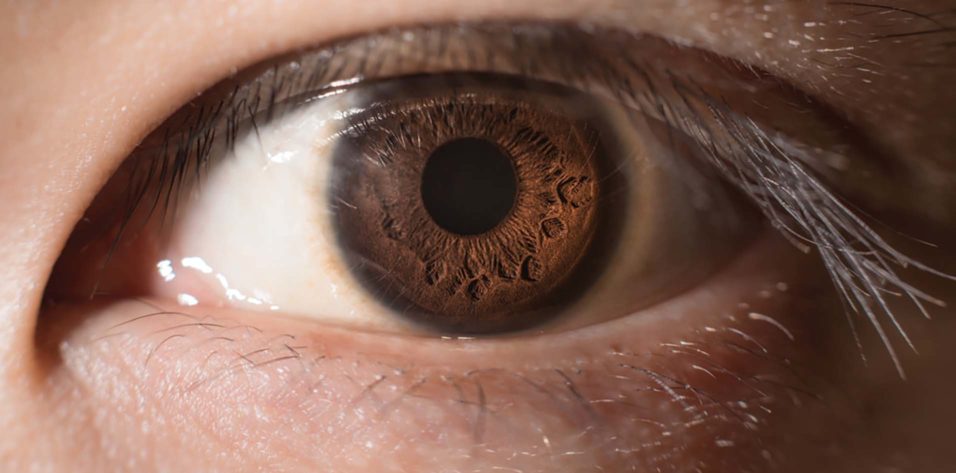
Normal pupil size in adults usually ranges from about 2 to 4 mm in bright light and 4 to 8 mm in the dark.
The exact value depends on light level, age, near effort and alertness, so a single number means little without the measurement conditions.
What Controls Normal Pupil Size
The iris sphincter constricts the pupil through parasympathetic fibers that travel with the third nerve, while the dilator muscle widens it under sympathetic control. As a result, pupil diameter reflects the moment-to-moment balance between these two systems.
Normal Pupil Size in Bright and Dim Light
Most healthy adults show pupils of roughly 2 to 4 mm in bright light, and in darkness the same pupils commonly dilate to about 4 to 8 mm. However, published ranges differ because studies use different luminance levels and field sizes. For example, Watson and Yellott combined several data sets into one formula that also includes age.
All-fit Slit-Lamp Adapter
Record and share exactly what you see at the slit lamp. One adapter fits any slit lamp or surgical microscope — and any smartphone.
From Choroida — the team behind this siteHow Normal Pupil Size Changes With Age
Newborns have small pupils, and diameter increases through childhood, with pupils tending to be largest in adolescence and early adulthood. After that, they decline steadily with age at every light level, and Winn and colleagues found this decline was linear and steepest in dim light.
Factors That Do Not Predict Normal Pupil Size
In the same study, sex, iris color and refractive error showed no significant relation to pupil size, so a blue-eyed myope should not be expected to have larger pupils. Wide individual variation persists, however, even among people of the same age.
Senile Miosis
Older patients often have small pupils that dilate poorly in the dark. This change likely reflects reduced sympathetic tone and age-related iris changes, and it lowers retinal illumination and may contribute to night vision complaints.
Physiologic Variation and Hippus
Healthy pupils are never perfectly still, and under steady light they show small rhythmic oscillations called hippus. In addition, pupils shrink with drowsiness and widen with arousal, fear or pain.
How to Measure Normal Pupil Size
First, ask the patient to fixate a distant target to avoid near miosis, and next measure each pupil in room light with a millimeter ruler or pupil gauge. Then dim the room and repeat, using faint light from below the chin.
Infrared pupillometers give the most precise readings, especially in dark irides, and finally, you should record the light condition beside every value.
Physiologic Anisocoria and Normal Pupil Size
A small difference between the two pupils is common in healthy people, and Lam, Thompson and Corbett found anisocoria of 0.4 mm or more in about one fifth of normal subjects. The difference usually stays under 1 mm and can vary from day to day, while both pupils react briskly and lids and motility are normal.
When Anisocoria Is Greater in the Dark
Anisocoria that increases in dim light suggests the smaller pupil dilates poorly. Horner syndrome is the main concern, especially with mild ptosis and dilation lag. Because physiologic anisocoria can behave similarly, apraclonidine or cocaine testing helps when suspicion is real.
When Anisocoria Is Greater in the Light
Anisocoria that increases in bright light suggests the larger pupil constricts poorly. The differential includes third nerve palsy, tonic pupil, iris trauma and pharmacologic mydriasis. For example, a tonic pupil shows segmental sphincter palsy and slow near constriction (see Adie tonic pupil).
Abnormally Large Pupils
A fixed dilated pupil with ptosis or diplopia needs urgent assessment for compressive third nerve palsy (see blown pupil). In contrast, an isolated dilated pupil with normal motility in a well patient is often pharmacologic, with scopolamine patches, plant alkaloids and nebulized ipratropium as typical sources.
Abnormally Small Pupils
Bilateral miosis commonly follows opioids or cholinergic drops such as pilocarpine, and posterior synechiae from uveitis can also fix a pupil in a small, irregular shape. Argyll Robertson pupils are small and irregular, with light-near dissociation (see Argyll Robertson pupil).
Normal Pupil Size Before Eye Surgery
Large dim-light pupils may increase halos and glare after some refractive and lens procedures, so surgeons often record scotopic diameter during planning. Small pupils, in contrast, can complicate phacoemulsification, especially with pseudoexfoliation or alpha-blocker use.
Counseling Patients About Normal Pupil Size
Patients sometimes notice unequal pupils in a mirror or photograph, and old photographs help date the finding and often confirm long-standing anisocoria. Explain that a small, stable difference with normal reactions is usually harmless. However, advise prompt review if new ptosis, double vision, headache or eye pain appears.
Pitfalls to Avoid
- Do not record a pupil diameter without noting the light condition
- Avoid measuring while the patient looks at a near target
- Never call anisocoria physiologic before checking lids, motility and light reactions
- Remember recent eye drops, scopolamine patches and nebulizers
- Do not compare readings from different devices as if they were equivalent


Document what you see
Two smartphone imaging tools built for everyday clinic use — one for the slit lamp, one for the fundus.
From Choroida — the team behind this siteReferences
- Winn B, Whitaker D, Elliott DB, Phillips NJ. Factors affecting light-adapted pupil size in normal human subjects. Invest Ophthalmol Vis Sci. 1994;35:1132-1137.
- Watson AB, Yellott JI. A unified formula for light-adapted pupil size. J Vis. 2012;12(10):12.
- Lam BL, Thompson HS, Corbett JJ. The prevalence of simple anisocoria. Am J Ophthalmol. 1987;104:69-73.
- Loewenfeld IE. The Pupil: Anatomy, Physiology, and Clinical Applications. 2nd ed. Boston: Butterworth-Heinemann; 1999.
- American Academy of Ophthalmology. Basic and Clinical Science Course, Section 5: Neuro-Ophthalmology. San Francisco: AAO; 2023.

Writes and reviews the clinical articles on Educate. Every post is checked against peer-reviewed sources before it goes live.