Optic nerve glioma is a low-grade astrocytic tumor of the anterior visual pathway that usually affects young children.
Most tumors are pilocytic astrocytomas, and many arise in neurofibromatosis type 1 (NF1). Because young children rarely report poor vision, the eye examination often gives the first clue.
Where Optic Nerve Glioma Grows
The tumor may involve one optic nerve, both nerves, the chiasm or the optic tracts, and for this reason clinicians often use the broader term optic pathway glioma. Orbital lesions cause proptosis and disc changes early. In contrast, chiasmal tumors cause bilateral field loss and endocrine signs.
Optic Nerve Glioma in NF1
Roughly 15 to 20 percent of children with NF1 show an optic pathway glioma on imaging, but only a minority ever lose vision. Most become apparent in early childhood, typically before age 7, and NF1 tumors also tend to behave more indolently than sporadic ones.
Fundus Explorer Pro
Photograph the retinal findings described here with the phone already in your pocket — 22 D optics and built-in illumination in one handheld unit.
From Choroida — the team behind this siteSporadic Optic Nerve Glioma
Children without NF1 more often present with chiasmal or hypothalamic disease, and these tumors may grow faster and threaten vision in both eyes. Then, hydrocephalus, nystagmus or precocious puberty may bring the child to attention first.
What the Patient Reports
Young children seldom complain, even with severe loss in one eye, so parents instead notice an eye turn, a head tilt, clumsiness or a bulging eye. Older children may describe blur, dim colors or a missing part of the field. As a result, history alone underestimates the disease.
Proptosis in Optic Nerve Glioma
Orbital tumors usually produce slowly progressive, painless axial proptosis, and the globe moves straight forward because the mass lies inside the muscle cone. In addition, large tumors can restrict motility or cause choroidal folds. Pain or rapid growth should therefore prompt a search for another diagnosis.
Optic Disc Changes
Optic atrophy is the most common fundus finding, and band or temporal pallor suggests chiasmal involvement (see temporal disc pallor). Disc swelling occurs less often, mainly with intraorbital tumors, and a normal disc does not exclude a tumor behind the globe.
Vision Loss in Optic Nerve Glioma
Visual acuity usually declines gradually and unevenly, and a relative afferent pupillary defect is a sensitive sign of asymmetric disease. However, symmetric bilateral involvement can leave the pupils equal, and color vision and fields add information, while reliable testing depends on age.
Eye Movement Signs
Poor vision in one eye often leads to sensory strabismus, and chiasmal tumors can also cause asymmetric nystagmus that mimics spasmus nutans. Therefore, every child with acquired nystagmus needs neuroimaging.
Screening for Optic Nerve Glioma in NF1
Consensus advice recommends a yearly comprehensive eye exam until about age 8, and after that, visits can space out through adolescence. Most experts advise against routine MRI in children without signs, because it rarely changes management.
The Screening Visit
- Age-appropriate visual acuity in each eye
- Pupil testing for a relative afferent defect
- Color vision and fields when feasible
- Exophthalmometry or inspection for proptosis
- Motility and nystagmus check
- Dilated fundus exam of disc color and contour
- Slit lamp search for Lisch nodules and lid neurofibroma
Imaging Optic Nerve Glioma
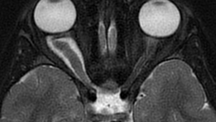
Contrast MRI of the brain and orbits is the study of choice, and NF1 tumors often show fusiform enlargement with kinking of the nerve. Some also show a perineural rim from arachnoidal gliomatosis, whereas sporadic tumors more often form a bulky chiasmal mass.
Differential Diagnosis
- Optic nerve sheath meningioma, mainly in adults (see optic nerve sheath meningioma)
- Optic neuritis, which is usually painful and subacute
- Craniopharyngioma or germinoma near the chiasm
- Orbital rhabdomyosarcoma, which grows rapidly
- Other causes of disc swelling (see optic disc swelling)
When to Treat Optic Nerve Glioma
Tumor size alone rarely triggers therapy, and instead most centers treat when acuity or fields decline on reliable testing, while growth on MRI with stable vision usually leads to closer follow-up.
Treatment Options
Carboplatin with vincristine is the usual first-line chemotherapy, which aims to stabilize vision, and some children improve. However, many keep their deficit, and some worsen despite treatment.
Oncologists increasingly use MEK or BRAF inhibitors for progressive or recurrent disease, while teams avoid radiation in young children with NF1 because of vasculopathy and secondary tumors. Surgery suits a blind, disfiguring eye or hydrocephalus.
Counseling Families
Explain that most NF1 gliomas never threaten sight, and stress that missed visits can delay detection of treatable loss. Treatment cannot restore atrophic axons, so earlier detection saves more vision.
Follow-Up
Children on treatment need regular acuity, field and fundus review, and nerve fiber layer OCT in addition gives objective data when fields are unreliable, while chiasmal disease also requires endocrine surveillance.
Pitfalls to Avoid
- Do not rely on symptoms in a preverbal child
- Do not test pupils in bright room light
- Never treat MRI growth alone without visual change
- Do not label unilateral nystagmus as spasmus nutans without imaging
- Avoid radiotherapy in NF1 when alternatives exist


Document what you see
Two smartphone imaging tools built for everyday clinic use — one for the slit lamp, one for the fundus.
From Choroida — the team behind this siteReferences
- Listernick R, Ferner RE, Liu GT, Gutmann DH. Optic pathway gliomas in neurofibromatosis-1: controversies and recommendations. Ann Neurol. 2007;61:189-198.
- Fisher MJ, Loguidice M, Gutmann DH, et al. Visual outcomes in children with neurofibromatosis type 1-associated optic pathway glioma following chemotherapy: a multicenter retrospective analysis. Neuro Oncol. 2012;14:790-797.
- Avery RA, Fisher MJ, Liu GT. Optic pathway gliomas. J Neuroophthalmol. 2011;31:269-278.
- Shapey J, Danesh-Meyer HV, Kaye AH. Diagnosis and management of optic nerve glioma. J Clin Neurosci. 2011;18:1585-1591.
- Listernick R, Charrow J, Greenwald M, Esterly NB. Optic gliomas in children with neurofibromatosis type 1. J Pediatr. 1989;114:788-792.
Test yourself
A few questions straight from this article.
-
What is the usual histological type of optic nerve glioma in children?
Optic nerve glioma is a low-grade astrocytic tumor of the anterior visual pathway, and most are pilocytic astrocytomas, many arising in NF1. -
Roughly what proportion of children with NF1 show an optic pathway glioma on imaging?
About 15 to 20 percent of NF1 children have an optic pathway glioma on imaging, yet only a minority ever lose vision. -
How do sporadic optic pathway gliomas typically differ from NF1-associated tumors?
Children without NF1 more often have chiasmal or hypothalamic tumors that may grow faster and threaten both eyes, while NF1 tumors behave more indolently. -
What type of proptosis does an intraorbital optic nerve glioma usually produce?
The mass lies inside the muscle cone, so the globe moves straight forward; pain or rapid growth should prompt a search for another diagnosis. -
What is the most common fundus finding in optic nerve glioma?
Optic atrophy is the most common finding, band or temporal pallor suggests chiasmal involvement, and disc swelling is less frequent. -
In optic nerve glioma, which sign is sensitive for asymmetric optic nerve involvement?
An RAPD is a sensitive sign of asymmetric disease, though symmetric bilateral involvement can leave the pupils equal. -
What screening is recommended for optic pathway glioma in asymptomatic children with NF1?
Consensus advice is a yearly eye exam until about age 8, and most experts advise against routine MRI in children without signs. -
What is the typical MRI appearance of an NF1-associated optic nerve glioma?
Contrast MRI of brain and orbits is the study of choice; NF1 tumors often show fusiform enlargement and kinking, whereas sporadic tumors form bulky chiasmal masses. -
What usually triggers treatment of an optic nerve glioma?
Most centers treat when vision declines on reliable testing; MRI growth with stable vision usually leads to closer follow-up instead. -
What is the usual first-line chemotherapy for a progressive optic pathway glioma?
Carboplatin with vincristine aims to stabilize vision; MEK or BRAF inhibitors are increasingly used for progressive disease, and radiation is avoided in young NF1 children.

Writes and reviews the clinical articles on Educate. Every post is checked against peer-reviewed sources before it goes live.