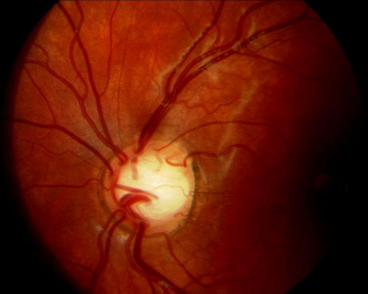
Megalopapilla is a congenitally large optic disc with a healthy neuroretinal rim and a large cup.
Because the cup looks big, these patients often arrive labeled as glaucoma suspects. However, the nerve tissue and its function are usually normal.
What Megalopapilla Is
Megalopapilla describes an optic disc with an area well above the normal range. Jonas and colleagues found wide variation in disc area among healthy eyes. Therefore, many authors call a disc large when its area exceeds about 2.5 square millimeters.
Why the Cup Is Large
Nerve fiber count varies much less than disc size. As a result, a wide scleral canal spreads a normal number of axons around a longer rim, and the rim stays normal in area while the empty center forms a big cup.
Fundus Explorer Pro
Photograph the retinal findings described here with the phone already in your pocket — 22 D optics and built-in illumination in one handheld unit.
From Choroida — the team behind this siteWho Has Large Discs
Disc size differs among ethnic groups. For example, people of African descent tend to have larger discs than White Europeans. High myopia can also enlarge the canal secondarily, and Jonas separated these acquired macrodiscs from the primary congenital form.
Symptoms in Megalopapilla
Most patients have no visual complaints, and acuity is usually normal. Therefore, the finding usually appears during routine fundus examination or glaucoma screening.
Fundus Signs of Megalopapilla
- Large disc relative to vessel caliber and disc-to-fovea distance
- Round or horizontally oval cup
- High cup-to-disc ratio, often symmetric in bilateral cases
- Pink rim without notches or focal thinning
- Rim width that follows the usual ISNT pattern
- No disc hemorrhage and no bayoneting of vessels
Why the Ratio Misleads
The cup-to-disc ratio ignores absolute disc size, so a 0.7 ratio in a large disc may contain more rim tissue than a 0.4 ratio in a small disc.
Garway-Heath and colleagues showed that the vertical ratio rises with disc area in normal eyes. Thus, interpret the ratio only after estimating disc size (see cup-to-disc ratio pitfalls).
Measuring Disc Size at the Slit Lamp
First, focus a 60, 78 or 90 diopter lens on the disc, and next narrow the beam and adjust its height to match the vertical disc diameter. Then read the beam height from the slit lamp scale.
Finally, apply the correction factor for that lens, because each lens magnifies differently. High refractive error also alters magnification, so treat the result as an estimate.
Interpreting the Measurement
Spaeth’s Disc Damage Likelihood Scale uses vertical diameter measured with a 60 diopter lens. With that lens, discs above 2.0 millimeters count as large, and the scale then grades the narrowest rim width according to disc size.
Megalopapilla on OCT
Optical coherence tomography defines the disc margin at Bruch’s membrane opening. In megalopapilla, retinal nerve fiber layer thickness and minimum rim width usually fall within normal limits. However, cup parameters may appear abnormal, so check the disc area before reading the color codes.
Visual Fields in Megalopapilla
Perimetry is usually normal, apart from a slightly enlarged blind spot. Therefore, an arcuate defect or nasal step needs another explanation, and a full, stable field over time supports the benign diagnosis.
Megalopapilla Versus Glaucoma
Glaucoma thins the rim focally and progressively. In contrast, megalopapilla keeps a uniform rim that does not change over time.
Notching, asymmetric cupping and raised pressure all shift the balance toward glaucoma, and a splinter hemorrhage at the margin signals active damage (see optic disc hemorrhage).
Other Large-Disc Mimics
Morning glory anomaly and optic disc coloboma also produce a large, excavated disc. However, they show glial tissue, abnormal vessels or inferior excavation, and they often reduce vision (see optic disc coloboma).
Can Glaucoma Coexist?
A large disc offers no protection against glaucoma. Therefore, a patient with megalopapilla can still develop glaucomatous damage later, and baseline photographs, OCT and fields make that change easier to detect.
Follow-Up in Megalopapilla
Many clinicians first repeat testing once or twice to confirm a stable rim and field. Afterward, routine eye examinations usually suffice when pressure stays normal. However, shorten the interval when a family history of glaucoma or other risk factors exist.
Counseling the Patient
Explain that the optic nerve is simply larger than average and looks healthy, and also ask the patient to mention the large disc at future eye visits.
Treatment in Megalopapilla
Megalopapilla itself needs no treatment. Glaucoma drops add cost and side effects without benefit when the nerve is healthy, so reserve treatment for documented glaucomatous damage or progression.
Pitfalls to Avoid
- Diagnosing glaucoma from the cup-to-disc ratio alone
- Grading the cup before estimating disc size
- Ignoring a notch or hemorrhage because the disc is large
- Trusting OCT color codes without checking disc area
- Reassuring a patient after one borderline visit


Document what you see
Two smartphone imaging tools built for everyday clinic use — one for the slit lamp, one for the fundus.
From Choroida — the team behind this siteReferences
- Jonas JB, Gusek GC, Naumann GO. Optic disc, cup and neuroretinal rim size, configuration and correlations in normal eyes. Invest Ophthalmol Vis Sci. 1988;29:1151-1158.
- Garway-Heath DF, Ruben ST, Viswanathan A, Hitchings RA. Vertical cup/disc ratio in relation to optic disc size: its value in the assessment of the glaucoma suspect. Br J Ophthalmol. 1998;82:1118-1124.
- Spaeth GL, Henderer J, Liu C, et al. The disc damage likelihood scale: reproducibility of a new method of estimating the amount of optic nerve damage caused by glaucoma. Trans Am Ophthalmol Soc. 2002;100:181-185.
- Brodsky MC. Congenital optic disk anomalies. Surv Ophthalmol. 1994;39:89-112.
Test yourself
A few questions straight from this article.
-
Which description defines megalopapilla?
Megalopapilla is a congenitally large optic disc whose neuroretinal rim is healthy; the cup looks big, but nerve tissue and function are usually normal. -
Why does megalopapilla produce a large optic cup despite healthy nerve tissue?
Nerve fiber count varies far less than disc size, so a wide scleral canal spreads a normal number of axons around a longer rim, leaving a large empty center. -
Above roughly what optic disc area do many authors call a disc large in megalopapilla?
Because disc area varies widely in healthy eyes, many authors call a disc large when its area exceeds about 2.5 square millimeters. -
On Spaeth's Disc Damage Likelihood Scale with a 60 diopter lens, which vertical disc diameter counts as large?
The DDLS uses vertical diameter measured with a 60 D lens; discs above 2.0 mm count as large, and rim width is then graded according to disc size. -
Why does the cup-to-disc ratio mislead in an eye with megalopapilla?
The ratio ignores disc size, so a 0.7 ratio in a large disc may contain more rim tissue than a 0.4 ratio in a small disc. -
What visual field finding is typical of megalopapilla?
Perimetry is usually normal apart from a slightly enlarged blind spot, so an arcuate defect or nasal step needs another explanation. -
Which rim behaviour distinguishes megalopapilla from glaucoma?
Glaucoma thins the rim focally and progressively, whereas megalopapilla keeps a uniform, stable rim; notching, asymmetric cupping and raised pressure point toward glaucoma. -
Which features separate morning glory anomaly and optic disc coloboma from megalopapilla?
These anomalies also give a large excavated disc but show glial tissue, abnormal vessels or inferior excavation, and they often reduce vision. -
What treatment does megalopapilla with a healthy optic nerve require?
Megalopapilla itself needs no treatment; glaucoma drops add cost and side effects without benefit, so treatment is reserved for documented damage or progression. -
On optic nerve OCT in megalopapilla, which parameters usually stay within normal limits?
RNFL thickness and minimum rim width usually fall within normal limits, but cup parameters may look abnormal, so check disc area before trusting the colour codes.

Writes and reviews the clinical articles on Educate. Every post is checked against peer-reviewed sources before it goes live.