Telecanthus is an increased distance between the medial canthi, while the distance between the pupils stays normal.
The bony orbits sit in their usual position, but the inner canthal soft tissue lies too far apart. Hypertelorism looks similar; however, it reflects widely separated bony orbits, and therefore a ruler and a careful exam usually separate the two.
What Telecanthus Means
The term describes lateral displacement of the medial canthal tendons, and as a result the inner corners look wide and the nasal bridge looks broad. The globes and pupils, however, keep their normal spacing, and because telecanthus is a sign, you should always look for its cause.
How Hypertelorism Differs
Hypertelorism means the medial orbital walls lie too far apart, and because the whole orbit shifts, the inner canthi, pupils and outer canthi all widen together. Consequently, intercanthal and interpupillary distances both rise, but only imaging measures the interorbital distance precisely.
All-fit Slit-Lamp Adapter
Record and share exactly what you see at the slit lamp. One adapter fits any slit lamp or surgical microscope — and any smartphone.
From Choroida — the team behind this siteMeasuring Intercanthal Distance
First, seat the patient facing you with the eyes in primary gaze, then measure between the medial canthal angles with a millimeter ruler. Avoid pressing on the skin because pressure shifts the soft tissue, and also record the palpebral fissure length.
Interpupillary and Outer Canthal Distance
Next, measure the interpupillary distance at distance fixation, and after that measure the outer canthal distance between the lateral canthi. Normal values vary with age, sex and ethnicity, so compare against published normograms, and in children, serial measurements help more than a single reading.
Ratios in Telecanthus
A common rule of thumb compares the intercanthal distance with half the interpupillary distance, and in most adults these two values are similar. When the intercanthal distance clearly exceeds that value, telecanthus is likely; however, if both distances rise together, suspect hypertelorism.
Pseudotelecanthus and Epicanthal Folds
Many infants look wide-eyed because of a flat nasal bridge and epicanthal folds, but in this case the measured intercanthal distance is normal. Pinching the nasal bridge lifts the folds and reveals the true canthal position, and the appearance usually improves as the face grows.
Primary Telecanthus in BPES
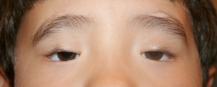
Blepharophimosis, ptosis, epicanthus inversus syndrome (BPES) is the classic congenital cause, and patients also have short fissures and an inverse epicanthal fold arising from the lower lid. FOXL2 mutations cause it, and type I carries a risk of premature ovarian insufficiency (see blepharophimosis syndrome). Therefore, examine the whole lid complex in any ptotic infant with telecanthus.
Waardenburg Syndrome and Dystopia Canthorum
Waardenburg syndrome type 1 displaces the inner canthi laterally, a sign called dystopia canthorum, and in addition patients may show heterochromia, a white forelock and hearing loss. The lacrimal puncta also shift laterally, which you can confirm at the slit lamp, and a biometric index helps separate type 1 from type 2.
Traumatic Telecanthus
Naso-orbito-ethmoidal fractures are the commonest acquired cause, and the medial canthal tendon may avulse or stay attached to a displaced central fragment. As a result, the canthal angle rounds and drifts laterally, and since globe injury often coexists, grade it carefully (see ocular trauma score).
Examining the Medial Canthal Tendon
The bowstring test checks tendon stability, so first pull the lower lid laterally while palpating the medial canthus. A firm endpoint suggests an intact tendon, while a soft canthus suggests detachment, and you should also test lacrimal drainage because canalicular injury often accompanies these fractures.
Causes of Hypertelorism
Hypertelorism usually reflects a craniofacial malformation, with examples including frontonasal dysplasia, facial clefts, and Crouzon or Apert syndrome. Therefore, these children need a craniofacial team, while the ophthalmologist monitors vision, exposure and strabismus (see exposure keratopathy).
Imaging in Telecanthus
Isolated congenital telecanthus rarely needs imaging; however, CT is the test of choice after facial trauma or when hypertelorism is suspected. Axial and coronal cuts show the medial orbital walls, bone fragments and interorbital distance, and the surgeon then plans repair from these images.
Pseudostrabismus in Telecanthus
Wide or folded inner corners hide the nasal sclera, and consequently parents often think the child has a turned eye. Check the corneal light reflex and perform a cover test before reassuring them, because true esotropia can coexist and you should never judge on appearance alone.
Surgical Repair of Telecanthus
Surgery moves the medial canthi nasally and reduces the epicanthal folds, and in BPES, surgeons often combine a Mustardé repair with transnasal canthopexy. Then ptosis repair follows, timed according to amblyopia risk, while traumatic cases need transnasal wiring or fixation of the central fragment, ideally early, although scarring, webbing and recurrence remain possible.
Counseling Families About Telecanthus
Explain that the eyes usually sit and work normally, and next discuss amblyopia, ptosis and refractive error, because these threaten vision most. For syndromic cases, offer genetic counseling and systemic referrals, and finally separate cosmetic goals from visual goals when timing surgery.
Pitfalls to Avoid
- Do not diagnose hypertelorism from soft tissue width alone
- Never skip the pinch test in infants with epicanthal folds
- Do not overlook the ovarian implications of BPES
- Always check the lacrimal system after naso-orbital trauma
- Do not label pseudostrabismus without a cover test


Document what you see
Two smartphone imaging tools built for everyday clinic use — one for the slit lamp, one for the fundus.
From Choroida — the team behind this siteReferences
- Crisponi L, Deiana M, Loi A, et al. The putative forkhead transcription factor FOXL2 is mutated in blepharophimosis/ptosis/epicanthus inversus syndrome. Nat Genet. 2001;27:159-166.
- Read AP, Newton VE. Waardenburg syndrome. J Med Genet. 1997;34:656-665.
- Markowitz BL, Manson PN, Sargent L, et al. Management of the medial canthal tendon in nasoethmoid orbital fractures. Plast Reconstr Surg. 1991;87:843-853.
- American Academy of Ophthalmology. Basic and Clinical Science Course, Section 7: Oculofacial Plastic and Orbital Surgery. San Francisco: AAO; 2023.

Writes and reviews the clinical articles on Educate. Every post is checked against peer-reviewed sources before it goes live.