Ocular hypotony is an intraocular pressure low enough to threaten the structure or function of the eye.
Statistically, a pressure below 6.5 mmHg lies more than three standard deviations under the population mean, but clinical significance depends on consequences, because some eyes tolerate low readings well.
Therefore, the workup asks why the pressure is low and whether the eye is being harmed.
Defining Ocular Hypotony
Many surgical studies use 5 mmHg or less as a working cutoff, while clinical hypotony means low pressure with visual loss, maculopathy, effusion or corneal folds.
Young myopes with thin sclera develop folds at pressures that older eyes ignore, so a single number should not drive treatment.
Two Mechanisms Behind Low Pressure
Every case reflects reduced aqueous production, increased outflow, or both, and outflow causes include leaks, overfiltration, cyclodialysis and retinal detachment.
In addition, production fails with ciliary inflammation, detachment, ischemia or destruction. After trauma, for example, a cleft and uveitis often act together.
All-fit Slit-Lamp Adapter
Record and share exactly what you see at the slit lamp. One adapter fits any slit lamp or surgical microscope — and any smartphone.
From Choroida — the team behind this siteCauses Checklist for Ocular Hypotony
- Wound leak after cataract, corneal or filtering surgery
- Overfiltration from a bleb or drainage device
- Cyclodialysis cleft after blunt trauma or surgery
- Ciliochoroidal detachment
- Rhegmatogenous retinal detachment, especially with proliferative vitreoretinopathy
- Uveitis with ciliary body shutdown
- Open globe injury, including occult posterior rupture
- Cyclodestruction and ocular ischemia
- Systemic causes such as dehydration, ketoacidosis or carbonic anhydrase inhibitors
History in Ocular Hypotony
First, establish the timeline of surgery, trauma and inflammation, and ask about filtering surgery, cataract incisions, intravitreal injections and blunt injury.
Also review medications and systemic illness, because bilateral hypotony points away from local causes and toward systemic disease.
Slit Lamp Signs
Look for a shallow chamber, Descemet folds, cells and flare, then inspect every wound and bleb for thinning and active drainage.
A diffuse, elevated bleb with low pressure suggests overfiltration. In contrast, a flat bleb with a shallow chamber suggests a leak or ciliary shutdown.
The Seidel Test in Ocular Hypotony
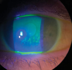
Paint concentrated fluorescein over the suspected site and examine under cobalt blue light, because aqueous dilutes the dye into a bright green stream that confirms an active leak.
In addition, gentle pressure on the globe can unmask a slow leak. However, a negative test cannot exclude an intermittent leak or posterior rupture.
Finding a Cyclodialysis Cleft
Gonioscopy shows a cleft as a deep gap between the scleral spur and ciliary body, but a shallow hypotonous chamber often hides it.
Ultrasound biomicroscopy or anterior segment OCT can then localize the cleft (see cyclodialysis cleft evaluation).
Fundus Examination in Ocular Hypotony
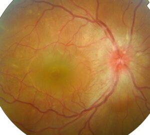
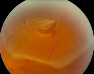
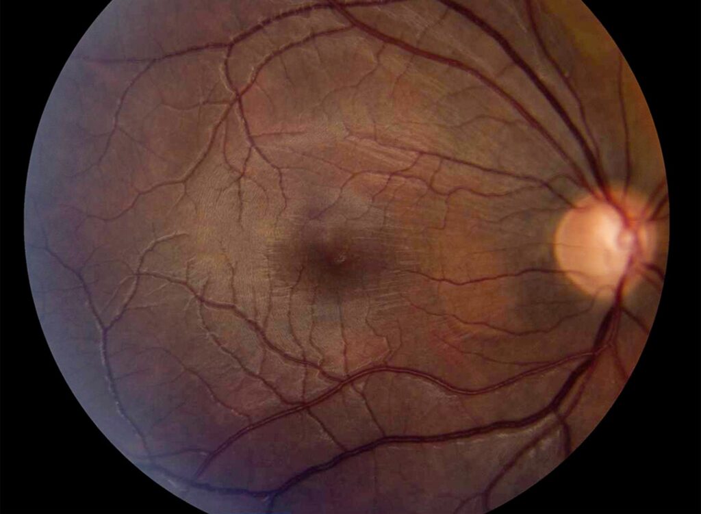
Dilated examination looks for choroidal folds, disc edema, venous tortuosity and retinal detachment, and smooth, dome-shaped peripheral elevations indicate choroidal effusion.
Also scan the periphery for breaks, because chronic detachment lowers pressure. Folds radiating through the fovea define hypotony maculopathy (see hypotony maculopathy management).
Imaging in Ocular Hypotony
B-scan ultrasound separates serous effusion from hemorrhage when media are opaque, and it also detects retinal detachment and posterior scleral rupture.
Meanwhile, OCT documents macular folds and tracks recovery.
Managing a Wound Leak
Small leaks often seal with a bandage contact lens and aqueous suppressants, and tapering topical steroids also helps, because they delay wound healing.
Persistent or large leaks need suturing, while leaking blebs may need revision.
Overfiltration After Glaucoma Surgery
Early hypotony after trabeculectomy often settles as the bleb matures, and next options include compression sutures, autologous blood injection and bleb revision.
After nonvalved tube surgery, ligation or flow restriction may be needed. The Tube Versus Trabeculectomy study reported persistent hypotony in both arms.
Closing a Cleft in Ocular Hypotony
Small clefts may close with cycloplegia, which relaxes the ciliary body toward the sclera, while larger or persistent clefts need laser, cryotherapy or direct cyclopexy.
Closure often causes a sharp pressure spike, so warn patients about pain and check pressure early.
Uveitis and Ciliary Shutdown
Active inflammation suppresses aqueous secretion by the ciliary epithelium, and therefore intensive steroid therapy often restores pressure.
Chronic disease, however, can cause cyclitic membranes and ciliary atrophy, so recovery is less likely in those eyes even after membrane removal.
Choroidal and Retinal Detachment
Serous choroidal detachment both results from hypotony and worsens it, and surgeons consider drainage for kissing choroids or lens-corneal touch (see choroidal detachment).
Rhegmatogenous detachment diverts fluid through the break toward the pigment epithelium, so successful repair often restores pressure.
Counseling and Follow-Up in Ocular Hypotony
Explain that low pressure blurs vision through folds, edema and choroidal fluid, and that many causes reverse with early treatment.
However, chronic hypotony can cause permanent maculopathy and eventually phthisis. Then recheck pressure, chamber depth and fundus at short intervals until stable.
Pitfalls to Avoid
- Do not treat a number without looking for structural damage
- Never stop at a negative Seidel test after suspicious trauma
- Do not miss a cleft hidden by a shallow chamber
- Remember systemic causes in bilateral hypotony


Document what you see
Two smartphone imaging tools built for everyday clinic use — one for the slit lamp, one for the fundus.
From Choroida — the team behind this siteReferences
- Wang Q, Thau A, Levin AV, Lee D. Ocular hypotony: a comprehensive review. Surv Ophthalmol. 2019;64:619-638.
- Pederson JE. Ocular hypotony. Trans Ophthalmol Soc U K. 1986;105:220-226.
- Costa VP, Arcieri ES. Hypotony maculopathy. Acta Ophthalmol Scand. 2007;85:586-597.
- Ioannidis AS, Barton K. Cyclodialysis cleft: causes and repair. Curr Opin Ophthalmol. 2010;21:150-154.
- Gedde SJ, Schiffman JC, Feuer WJ, et al. Treatment outcomes in the Tube Versus Trabeculectomy (TVT) study after five years of follow-up. Am J Ophthalmol. 2012;153:789-803.

Writes and reviews the clinical articles on Educate. Every post is checked against peer-reviewed sources before it goes live.