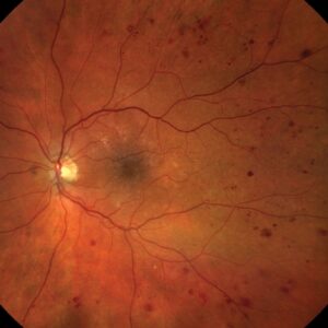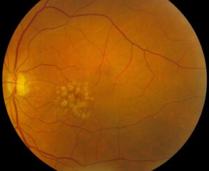
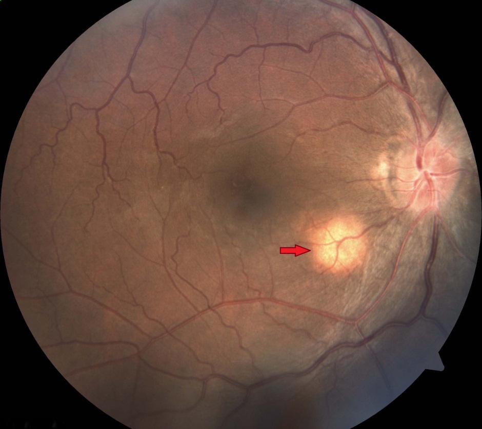
Choroidal granuloma is a focal inflammatory mass in the choroid, usually caused by tuberculosis, sarcoidosis or fungal infection.
At first glance, it can look exactly like a tumor. Therefore, the workup must separate infection, systemic inflammation and neoplasm before any steroid starts.
What a Choroidal Granuloma Is
A granuloma is an organized collection of macrophages, epithelioid cells and lymphocytes, and in the choroid it forms an elevated nodule beneath the retinal pigment epithelium. However, a large solitary mass raises the hardest diagnostic questions.
Causes of Choroidal Granuloma
- Tuberculosis, the classic solitary mass in endemic regions
- Sarcoidosis, often with multiple yellow nodules
- Fungi, for example Cryptococcus or Candida in immunocompromised patients
- Syphilis, which can mimic almost any posterior lesion
- Toxocara, which forms a posterior pole granuloma in children
Fundus Explorer Pro
Photograph the retinal findings described here with the phone already in your pocket — 22 D optics and built-in illumination in one handheld unit.
From Choroida — the team behind this siteTuberculoma: The Classic Solitary Lesion
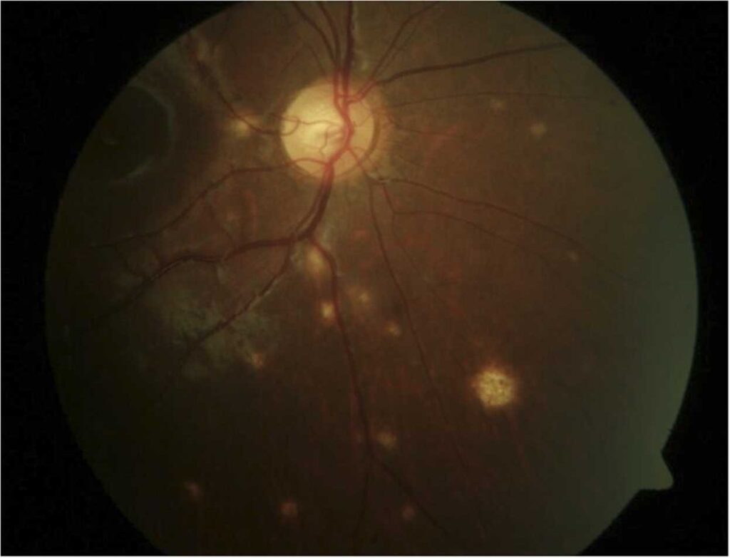
Choroidal tuberculoma usually appears as a yellow or gray-white elevated mass, and it often sits at the posterior pole, sometimes with overlying exudative retinal detachment.
In addition, vitritis and anterior chamber cells are common, although some eyes stay quiet. A positive interferon-gamma release assay supports the diagnosis but does not prove it.
Sarcoid Choroidal Granuloma
Sarcoidosis tends to produce multiple small yellow choroidal nodules. However, a solitary large granuloma can occur and may mimic metastasis.
Also, look for mutton-fat keratic precipitates, iris nodules and periphlebitis (see sarcoid uveitis). Chest imaging and serum markers then guide the systemic search.
Fungal Choroidal Granuloma
Fungal lesions appear mainly in immunocompromised patients, intravenous drug users or after candidemia, and cryptococcal choroiditis may present as one or several cream-colored lesions.
The main danger is empirical steroid therapy, because it can accelerate infection. Therefore, ask about immune status, indwelling lines and recent hospital admission.
OCT in Choroidal Granuloma
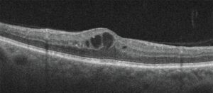
Spectral-domain OCT shows a dome-shaped elevation with loss of the normal choroidal vascular pattern. Over the lesion, a focal adhesion between the RPE and outer retina has been described as a “contact sign”.
Subretinal fluid often surrounds this contact zone, and enhanced depth imaging then provides a thickness baseline for follow-up.
Angiography and Ultrasound
On fluorescein angiography, active granulomas often show early hypofluorescence and late leakage, while indocyanine green angiography typically shows dark lesions that persist into late phases.
Meanwhile, B-scan confirms a solid mass and measures its height. However, these patterns overlap with tumors, so no single test settles the question.
Choroidal Granuloma Versus Tumor
The main mimics are amelanotic melanoma, metastasis and lymphoma. Melanoma grows slowly and may show a collar-button shape on ultrasound (see choroidal melanoma).
Metastases are often multiple, with a known primary in many patients (see choroidal metastases). In contrast, granulomas usually come with vitritis and change within weeks.
Laboratory Workup in Choroidal Granuloma
Start with an interferon-gamma release assay or tuberculin skin test, plus chest imaging. Next, order treponemal and nontreponemal syphilis serology.
Serum angiotensin-converting enzyme and lysozyme help when sarcoidosis is suspected. Finally, add HIV testing and blood cultures when fungal disease is possible.
Treatment of Tubercular Choroidal Granuloma
Antitubercular therapy, coordinated with an infectious disease physician, treats the cause, and oral corticosteroids are often added once therapy is under way to limit tissue damage.
Collaborative cohort data show variable responses, so case selection deserves care. Also, paradoxical worsening can occur early and does not mean treatment failure.
Treatment for Sarcoid and Fungal Lesions
Sarcoid granulomas usually respond to corticosteroids, given locally or systemically, and when long courses are needed, steroid-sparing immunosuppression follows.
Fungal granulomas, by contrast, need systemic antifungal therapy, sometimes with intravitreal injection. Drug choice and duration then follow infectious disease advice.
How Treatment Response Confirms Choroidal Granuloma
Inflammatory masses usually flatten and scar within weeks of correct therapy. As a result, measured regression supports the working diagnosis.
Melanoma and metastasis do not regress with antitubercular drugs. Therefore, document size with photos, OCT and ultrasound before treatment begins.
When to Biopsy a Choroidal Granuloma
Biopsy becomes reasonable when the lesion grows despite therapy or the workup stays negative. Fine-needle aspiration or vitrectomy-based biopsy can provide cytology, culture and PCR. Discuss bleeding, retinal detachment and nondiagnostic samples with the patient beforehand.
Counseling the Patient
Explain that the mass is probably inflammatory, yet tumor stays on the list until it responds. Then outline the treatment plan, expected timeline and follow-up schedule. Patients on long antitubercular courses need support with adherence and side effects.
Pitfalls to Avoid
- Do not start steroids alone before excluding tuberculosis, syphilis and fungi
- Never label a solitary amelanotic mass inflammatory without baseline imaging
- Do not stop antitubercular therapy at the first paradoxical flare
- Remember that steroid response does not exclude lymphoma


Document what you see
Two smartphone imaging tools built for everyday clinic use — one for the slit lamp, one for the fundus.
From Choroida — the team behind this siteReferences
- Gupta V, Gupta A, Rao NA. Intraocular tuberculosis: an update. Surv Ophthalmol. 2007;52:561-587.
- Herbort CP, Rao NA, Mochizuki M, et al. International criteria for the diagnosis of ocular sarcoidosis. Ocul Immunol Inflamm. 2009;17:160-169.
- Agrawal R, Gunasekeran DV, Grant R, et al. Clinical features and outcomes of patients with tubercular uveitis treated with antitubercular therapy in the Collaborative Ocular Tuberculosis Study (COTS)-1. JAMA Ophthalmol. 2017;135:1318-1327.
- Shields JA, Mashayekhi A, Ra S, Shields CL. Pseudomelanomas of the posterior uveal tract: the 2006 Taylor R. Smith lecture. Retina. 2005;25:767-771.
- Salmon JF. Kanski’s Clinical Ophthalmology: A Systematic Approach. 9th ed. Elsevier; 2020.
Test yourself
A few questions straight from this article.
-
What cells make up a choroidal granuloma?
A granuloma is an organized collection of macrophages, epithelioid cells and lymphocytes, forming an elevated nodule beneath the RPE in the choroid. -
Which cause classically produces a solitary choroidal granuloma in endemic regions?
Tuberculosis is the classic solitary mass in endemic regions, often a yellow or gray-white posterior pole lesion, whereas sarcoidosis tends to produce multiple small yellow nodules. -
Which anterior segment findings support sarcoidosis as the cause of a choroidal granuloma?
With a suspected sarcoid granuloma, mutton-fat keratic precipitates, iris nodules and periphlebitis are sought, and chest imaging and serum markers guide the systemic search. -
In a suspected fungal choroidal granuloma, what is the main danger?
Fungal lesions occur mainly in immunocompromised patients, IV drug users or after candidemia, and empirical steroids can accelerate the infection. -
On OCT of a choroidal granuloma, what is the 'contact sign'?
SD-OCT shows a dome-shaped elevation with loss of the choroidal vascular pattern, and a focal RPE-outer retina adhesion over it has been called the contact sign, often surrounded by subretinal fluid. -
Which ultrasound shape favours amelanotic melanoma over a choroidal granuloma?
Melanoma grows slowly and may show a collar-button shape on ultrasound, whereas granulomas usually come with vitritis and change within weeks. -
In suspected choroidal tuberculoma, what does a positive interferon-gamma release assay mean?
A positive interferon-gamma release assay supports the diagnosis of choroidal tuberculoma but does not prove it, so the full workup still proceeds. -
A tubercular choroidal granuloma worsens early after antitubercular therapy starts. How should this be interpreted?
Paradoxical worsening can occur early in antitubercular therapy and does not mean treatment failure, so therapy should not be stopped at the first flare. -
Why should a choroidal granuloma's size be documented with photos, OCT and ultrasound before treatment?
Inflammatory masses usually flatten and scar within weeks of correct therapy, while melanoma and metastasis do not regress with antitubercular drugs, so measured regression supports the diagnosis. -
When does biopsy of a presumed choroidal granuloma become reasonable?
Biopsy by fine-needle aspiration or vitrectomy is reasonable when the lesion grows despite therapy or the workup stays negative, after discussing bleeding, detachment and nondiagnostic samples.

Writes and reviews the clinical articles on Educate. Every post is checked against peer-reviewed sources before it goes live.