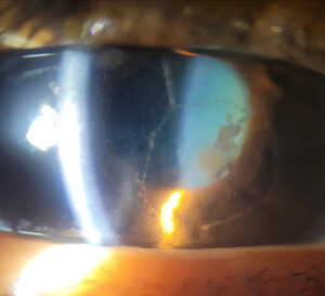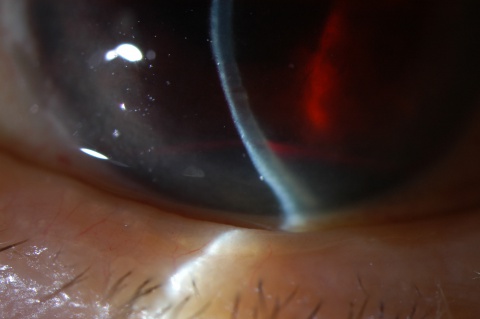
Microhyphema means red blood cells circulating in the anterior chamber without a layered clot.
It usually follows blunt trauma, and a penlight examination can easily miss it.
However, it can still cause rebleeding and raised intraocular pressure (IOP).
Therefore, an injured eye with blurred vision needs a slit lamp examination.
What Microhyphema Means
Trauma can tear vessels in the iris or ciliary body, releasing red cells into the aqueous.
In a layered hyphema, those cells settle into a visible inferior meniscus.
In microhyphema, they stay suspended, so the chamber may look clear on gross inspection.
Many grading schemes label this finding grade 0.
Symptoms and History
Patients often describe blurred vision, photophobia or a dull ache after an injury.
However, vision can remain good, so normal acuity does not exclude the diagnosis.
Also, ask about anticoagulants, antiplatelet drugs, bleeding disorders and sickle status.
All-fit Slit-Lamp Adapter
Record and share exactly what you see at the slit lamp. One adapter fits any slit lamp or surgical microscope — and any smartphone.
From Choroida — the team behind this siteSlit Lamp Technique for Microhyphema
First, dim the room and allow your eyes to adapt.
Then, set a short, bright oblique beam about 1 mm long.
Next, use high magnification and focus on the space between cornea and lens.
Finally, watch for cells drifting in the convection current.
Recognizing Red Cells
Red cells appear small and reddish, while white cells look gray-white.
Pigment granules, by contrast, often look brown and irregular.
In addition, inspect the inferior chamber, because a thin layer changes the grade.
Why Microhyphema Can Layer Later
Red cells sink with gravity, so an upright patient may develop a thin inferior layer.
Therefore, repeat the examination after upright rest when the first view seems equivocal.
However, rising cell density raises concern for fresh bleeding.
Causes Beyond Trauma
A spontaneous bleed needs a source, so never accept an atraumatic history without investigation.
Check the pupil margin and angle for neovascularization.
Also, consider uveitis-glaucoma-hyphema syndrome when an intraocular lens may be rubbing the iris.
The broader hyphema review covers these causes in more detail.
Differential Diagnosis
Anterior uveitis produces white cells and flare, often with keratic precipitates.
Meanwhile, khaki cells after vitreous hemorrhage suggest ghost cell glaucoma.
Therefore, record cell color as well as cell count.
Excluding Open Globe Injury
A small bleed does not prove that the globe is intact.
Therefore, look for an irregular pupil, a shallow chamber or unexpected hypotony.
If rupture seems possible, stop contact tests and place a rigid shield.
Then, arrange urgent surgical assessment.
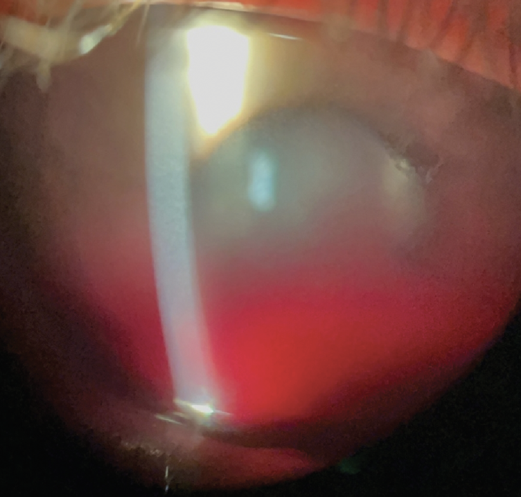
IOP in Microhyphema
Red cells can obstruct the trabecular meshwork, even without a visible clot.
As a result, IOP may rise at presentation or after a rebleed.
Therefore, measure pressure at every visit.
Sickle Status and Microhyphema
Sickled cells pass poorly through the outflow pathway, so modest bleeding can produce dangerous pressure.
Therefore, test patients at risk when sickle status is unknown.
Our article on sickle cell hyphema explains the lower treatment thresholds.
Treating Microhyphema
Treatment aims to limit rebleeding, control inflammation and protect the optic nerve.
A rigid shield, head elevation and reduced activity are common supportive measures.
Also, avoid nonessential aspirin and NSAIDs, while coordinating anticoagulant decisions with the responsible physician.
Topical steroids and cycloplegics may improve comfort.
For elevated IOP, topical aqueous suppressants are usually first choice.
However, avoid carbonic anhydrase inhibitors in sickle disease when possible.
What the Evidence Shows
A Cochrane review found no clear visual benefit from common medical interventions in traumatic hyphema.
Antifibrinolytic agents may reduce rebleeding, but they carry adverse effects.
Moreover, most trials did not focus on microhyphema.
Therefore, management relies partly on clinical judgment.
Outpatient Care Decisions
A retrospective series by Recchia and colleagues supported outpatient management in selected patients.
However, that design cannot define a universally safe pathway.
Reliable follow-up, controlled IOP and an intact globe favor home care.
Conversely, sickle disease or poor adherence may justify closer supervision.
Follow-Up of Microhyphema
Document cell density, cell color, IOP and any new inferior layer.
Rebleeding typically occurs within the first several days, so early visits should cover this period.
Then, extend intervals as cells clear.
Any new layer, pain or pressure rise should prompt earlier reassessment.
Late Examination After Injury
Once the bleed resolves, examine the angle and peripheral retina.
Gonioscopy can reveal angle recession, while dilated examination can detect retinal tears.
Later, angle recession calls for long-term IOP monitoring, because glaucoma can develop years afterward.
Counseling Patients with Microhyphema
Explain that a small bleed can still worsen during the first days.
Next, ask patients to report sudden vision loss, increasing pain or fresh redness.
Finally, recommend protective eyewear for future sports.
Pitfalls to Avoid
- Do not call the chamber clear after a penlight examination alone
- Do not label every traumatic cell as iritis without assessing cell color
- Never skip IOP measurement once open globe injury seems unlikely
- Do not overlook sickle testing in patients at risk


Document what you see
Two smartphone imaging tools built for everyday clinic use — one for the slit lamp, one for the fundus.
From Choroida — the team behind this siteReferences
- Walton W, Von Hagen S, Grigorian R, Zarbin M. Management of traumatic hyphema. Surv Ophthalmol. 2002;47:297-334.
- Gharaibeh A, Savage HI, Scherer RW, Goldberg MF, Lindsley K. Medical interventions for traumatic hyphema. Cochrane Database Syst Rev. 2019;1:CD005431.
- Recchia FM, Saluja RK, Hammel K, Jeffers JB. Outpatient management of traumatic microhyphema. Ophthalmology. 2002;109:1465-1470.
- Bansal S, Gunasekeran DV, Ang B, et al. Controversies in the pathophysiology and management of hyphema. Surv Ophthalmol. 2016;61:297-308.

Writes and reviews the clinical articles on Educate. Every post is checked against peer-reviewed sources before it goes live.