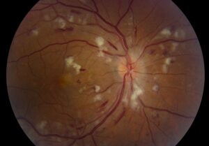
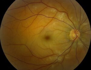
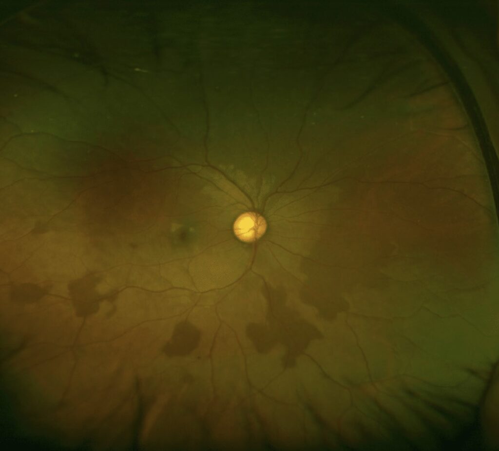
Dark without pressure is a flat, dark brown or gray patch in the peripheral retina.
It typically appears as an incidental finding during dilated examination.
Because its borders look sharp, it often prompts referral.
However, the lesion usually follows a benign course.
What Is Dark Without Pressure?
The examiner sees a region of retina darker than its surroundings without any scleral depression.
Its counterpart is white without pressure, a pale patch with similar geographic borders.
In addition, both findings may occur in the same eye.
Who Develops Dark Without Pressure?
Clinicians report this finding more often in younger patients and in darkly pigmented fundi.
Early work described these lesions in patients with sickle hemoglobinopathies.
However, otherwise healthy people also show them, so the finding alone does not establish a systemic diagnosis.
Fundus Explorer Pro
Photograph the retinal findings described here with the phone already in your pocket — 22 D optics and built-in illumination in one handheld unit.
From Choroida — the team behind this siteSymptoms and History
Most patients report no visual symptoms.
Therefore, do not attribute new flashes, floaters or a field defect to the patch.
Next, ask about sickle cell disease, trauma and prior retinal disease.
Exam Signs of Dark Without Pressure
Indirect ophthalmoscopy usually shows a recognizable pattern:
- Flat, geographic patch with sharp, often scalloped borders
- Dark brown or gray color, usually homogeneous
- Retinal vessels crossing the area without displacement
- No subretinal fluid, hemorrhage or orange pigment
Examination Technique
First, dilate the pupil fully and use indirect ophthalmoscopy.
Then, scan every quadrant, because the patch often lies near the ora serrata.
Next, use scleral depression to check the adjacent retina for breaks.
Also, compare the fellow eye before you reach a conclusion.
Likely Mechanism
The exact cause remains uncertain.
Multimodal imaging points toward altered reflectivity in the outer retina, particularly at the ellipsoid zone.
Some authors also suspect a vitreoretinal contribution, but that link remains a hypothesis.
OCT in Dark Without Pressure
Spectral-domain OCT can support the diagnosis when the lesion lies within scanning range.
Fawzi and colleagues described reduced ellipsoid zone reflectivity with an abrupt transition at the lesion border.
Meanwhile, the scan should show no solid mass and no subretinal fluid.
Peripheral lesions are hard to capture, so a limited scan does not exclude other disease.
Other Imaging Tests
Widefield color photography gives the most practical baseline record.
It also allows comparison of size and shape at later visits.
Fluorescein angiography is usually unnecessary.
However, consider it when you suspect sickle-related nonperfusion.
Natural History
These patches can change over time.
For example, they may enlarge, shrink, fade or appear to shift position.
As a result, a change in outline does not suggest malignancy when the lesion stays flat and typical.
Mimics to Exclude
Several dark lesions resemble this finding, and some need different management (see peripheral retinal variants):
- Congenital hypertrophy of the retinal pigment epithelium, often with lacunae
- Choroidal nevus, which has a deeper appearance and may show drusen
- Choroidal melanoma, which may show thickness, fluid or orange pigment
- Subretinal or sub-RPE hemorrhage
- Black sunburst lesions in sickle retinopathy
Separating Melanoma From Dark Without Pressure
Elevation is the first feature to assess.
Next, look for subretinal fluid, orange pigment and thickness on ultrasound.
Any of these features calls for prompt referral to ocular oncology.
A flat patch with crisp borders and normal overlying vessels points the other way.
Sickle Disease and the Patch
Routine hemoglobin testing is not required for every patient.
However, testing becomes reasonable with suggestive history, ancestry or associated fundus signs.
For example, look for peripheral nonperfusion, salmon patch hemorrhages and sea fan neovascularization.
Retinal Breaks and Detachment Risk
Current evidence does not establish this lesion as a precursor of retinal detachment.
Still, its presence does not exclude a separate peripheral problem.
Symptomatic patients therefore need a depressed examination for breaks (see rhegmatogenous retinal detachment).
Treatment of Dark Without Pressure
Typical lesions need no treatment.
Observational reports support this approach, and no controlled trials have tested an intervention.
Prophylactic laser has no established role, and it carries its own risks.
It also cannot remove the patch.
When to Refer
Refer promptly if the lesion is elevated, shows fluid or grows steadily.
Likewise, refer when vision falls without another explanation.
However, a flat, stable and typical patch rarely needs specialist review.
Follow-Up and Counseling
First, document the lesion with a drawing or widefield photograph.
Then, return a typical asymptomatic patient to routine dilated examinations.
Explain that the outline may vary between visits.
However, advise patients to report flashes, floaters, a curtain or reduced vision immediately.
Pitfalls to Avoid
- Do not label an elevated pigmented lesion as dark without pressure
- Avoid laser treatment for an isolated flat patch
- Do not skip scleral depression when symptoms suggest a break
- Do not ignore other signs of sickle retinopathy


Document what you see
Two smartphone imaging tools built for everyday clinic use — one for the slit lamp, one for the fundus.
From Choroida — the team behind this siteReferences
- Nagpal KC, Goldbaum M, Asdourian G, Goldberg MF, Huamonte F. Dark-without-pressure fundus lesions. Br J Ophthalmol. 1975;59:476-479.
- Fawzi AA, Nielsen JS, Mateo-Montoya A, et al. Multimodal imaging of white and dark without pressure fundus lesions. Retina. 2014;34:2376-2387.
- Shields CL, Mashayekhi A, Ho T, Cater J, Shields JA. Solitary congenital hypertrophy of the retinal pigment epithelium: clinical features and frequency of enlargement in 330 patients. Ophthalmology. 2003;110:1968-1976.
- Flaxel CJ, Adelman RA, Bailey ST, et al. Posterior vitreous detachment, retinal breaks, and lattice degeneration Preferred Practice Pattern. Ophthalmology. 2020;127:P146-P181.
Test yourself
A few questions straight from this article.
-
How is dark without pressure defined on peripheral retinal examination?
Dark without pressure is a flat, dark brown or gray area of retina darker than its surroundings, seen without any scleral depression; white without pressure is its pale counterpart. -
In which patients is dark without pressure reported more often?
Clinicians report dark without pressure more often in younger patients and in darkly pigmented fundi, and also in healthy people. -
Which indirect ophthalmoscopy finding is typical of dark without pressure?
The typical patch is flat and geographic with sharp, often scalloped borders, and retinal vessels cross it without displacement. -
What does spectral-domain OCT typically show at a dark without pressure lesion?
Fawzi and colleagues described reduced ellipsoid zone reflectivity with an abrupt transition at the lesion border, with no mass or subretinal fluid. -
A flat, typical dark without pressure patch changes outline between visits. How should this be interpreted?
These patches can enlarge, shrink, fade or seem to shift, so a change in outline does not suggest malignancy when the lesion stays flat and typical. -
When separating choroidal melanoma from dark without pressure, which feature should be assessed first?
Elevation is assessed first, then subretinal fluid, orange pigment and ultrasound thickness; any of these calls for prompt ocular oncology referral. -
Which mimic of dark without pressure often shows lacunae?
Congenital hypertrophy of the retinal pigment epithelium, often with lacunae, is one of the dark lesions to exclude, alongside nevus, melanoma, hemorrhage and black sunburst. -
What is the recommended treatment for a typical dark without pressure lesion?
Typical lesions need no treatment; prophylactic laser has no established role, carries its own risks and cannot remove the patch. -
When is hemoglobin testing for sickle disease reasonable in a patient with dark without pressure?
Routine hemoglobin testing is not required, but it becomes reasonable with suggestive history, ancestry or signs such as peripheral nonperfusion or sea fan neovascularization. -
A patient with known dark without pressure reports new flashes and floaters. What is the appropriate step?
The lesion is not established as a detachment precursor and should not explain new symptoms, so symptomatic patients need a depressed examination for breaks.

Writes and reviews the clinical articles on Educate. Every post is checked against peer-reviewed sources before it goes live.