The photostress test measures how long the macula takes to recover after a bright light bleaches its photopigment.
It needs only a penlight and a visual acuity chart, and it helps to separate macular disease from optic nerve disease.
The test is simple, but its interpretation needs care.
How the Photostress Test Works
A bright light bleaches the visual pigment in cone photoreceptors, and vision drops.
Recovery needs regeneration of the pigment, which depends on the health of the retinal pigment epithelium and the photoreceptors.
Macular disease slows this recovery, whereas optic nerve disease does not affect the pigment cycle.
How to Perform the Photostress Test
Measure the best-corrected acuity in the eye at baseline, and note the line.
Hold a bright penlight or ophthalmoscope light about 3 cm from the eye for 10 seconds, with the patient looking at the light.
Then ask the patient to read the line one level above the baseline line, and time how long recovery takes.
Repeat for the fellow eye and compare, since the two eyes should recover at similar rates.
Fundus Explorer Pro
Photograph the retinal findings described here with the phone already in your pocket — 22 D optics and built-in illumination in one handheld unit.
From Choroida — the team behind this siteNormal and Abnormal Photostress Test Results
Normal recovery usually takes less than 50 seconds, though values vary with the light source and the technique.
Glaser and colleagues reported that prolonged recovery times were found in macular disease and normal times in optic nerve disease.
Severin and colleagues linked recovery time to macular function in early studies (see toxic nutritional optic neuropathy).
Margrain and Thomson found significant variability from test to test, so repeat the measure when results are borderline.
Uses of the Photostress Test
- Unexplained loss of acuity with a normal fundus
- Early central serous disease, with a prolonged recovery
- Differentiating macular from optic nerve disease
- Following recovery after macular treatment
In optic neuropathy, recovery is usually normal, and a relative afferent defect and color loss are more informative.
Limits of the Photostress Test
The test is not specific for any disease, and it does not replace OCT or ERG.
Media opacity, uncorrected refractive error, and poor cooperation reduce its value.
Standardize the light distance, the duration, and the target line to improve repeatability.
Practical Points
Use the test early in the examination when a diagnosis is not obvious.
Document the baseline line, the recovery time, and the light source.
Then repeat the test after treatment to follow function.
An Example Case
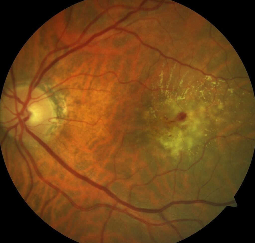
A 45-year-old woman has 20/40 vision in her right eye and a normal fundus.
The photostress recovery time is 90 seconds in the right eye and 25 seconds in the left eye.
OCT shows subtle subretinal fluid at the fovea, consistent with central serous disease.
After the fluid resolves, her acuity returns to 20/20, and the recovery time falls to 30 seconds.
Questions Patients Ask
Patients ask whether the bright light harms the eye, and a ten-second exposure with a penlight is safe.
They also ask why vision is blurry afterward, and that is the normal after-image from the bleached pigment.
Because the after-image can alarm some patients, explain it before the test.
Pitfalls to Avoid
- Using different lights from one visit to the next
- Letting the patient look away from the light during exposure
- Reading the result without comparing the fellow eye
- Treating a prolonged time as a diagnosis, not a clue
Looking Ahead
Objective tests such as pattern ERG and OCT have reduced the use of the photostress test.
Still, the test remains a fast office screen where equipment is limited.
Therefore, learn it as a clinical principle as well as a procedure.
Practical Points
Use a consistent light source and a fixed distance.
Then measure at the same baseline acuity line each time.
Also note whether the patient reports a central dark after-image.
Variants of the Test
Some clinicians use a direct ophthalmoscope for 10 seconds, and others use a flash from a camera.
Because the light source affects the result, keep your method constant.
Then build your own range for the method, since published values differ.
Interpreting Borderline Results
When recovery time is 50 to 60 seconds, repeat the test and compare with the other eye.
If the difference persists, order OCT and consider electrophysiology.
Also check for media opacities, since they slow recovery and may mislead.
Recording the Result
Write the baseline line, the light source, and the recovery time in seconds.
Then note the fellow eye value, since the difference is more useful than the absolute number.
Also repeat the test at follow-up under the same conditions.
For the latest studies, see the PubMed results for photostress test.


Document what you see
Two smartphone imaging tools built for everyday clinic use — one for the slit lamp, one for the fundus.
From Choroida — the team behind this siteReferences
- Glaser JS, Savino PJ, Sumers KD, et al. The photostress recovery test in the clinical assessment of visual function. Am J Ophthalmol. 1977;83:255-260.
- Severin SL, Tour RL, Kershaw RH. Macular function and the photostress test. Arch Ophthalmol. 1967;77:163-167.
- Margrain TH, Thomson D. Sources of variability in the clinical photostress test. Ophthalmic Physiol Opt. 2002;22:61-67.
- American Academy of Ophthalmology. Basic and Clinical Science Course, Section 12: Retina and Vitreous. AAO; current edition.

Writes and reviews the clinical articles on Educate. Every post is checked against peer-reviewed sources before it goes live.