
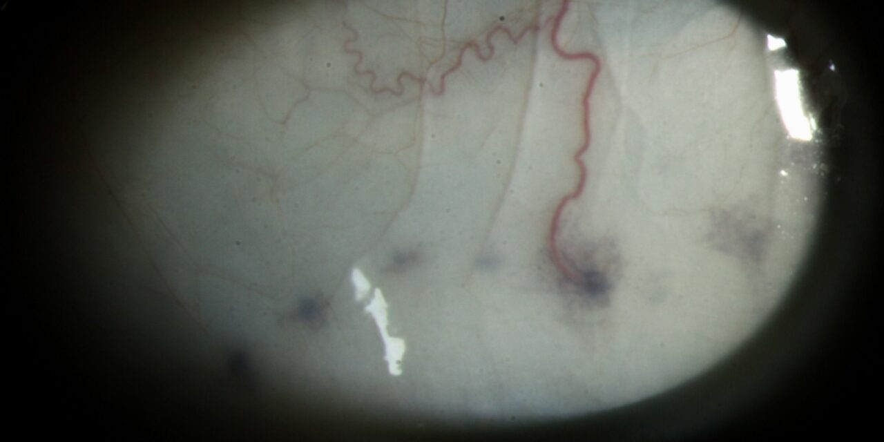
A patient notices a small dark spot on the white of the eye, and a doctor sees a pigmented bump a few millimeters behind the limbus.
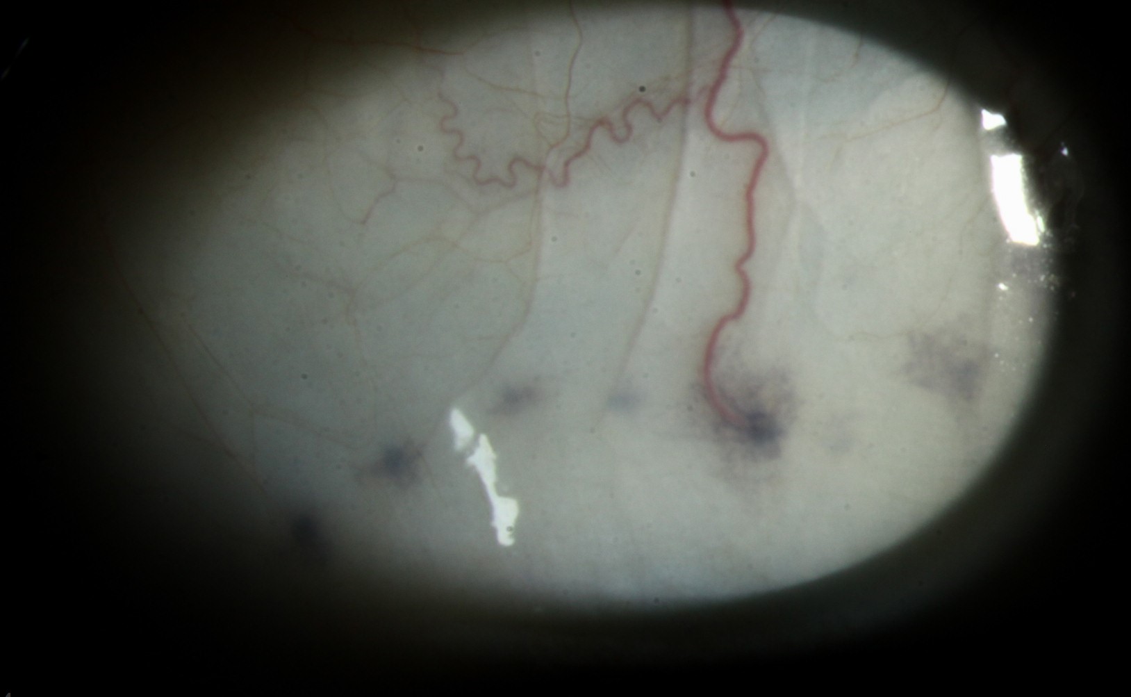
A melanoma is feared, and the patient is sent for evaluation.
In a fair number of those cases, the final diagnosis is an Axenfeld nerve loop, a normal anatomical variant.
What it is
A long posterior ciliary nerve runs forward from the optic nerve in the suprachoroidal space.
In some individuals, a loop of this nerve penetrates a short distance into the sclera before returning, and the thinned sclera over it lets the nerve and the pigment from the nearby uvea show through.
The result is a small, round or oval, gray-to-blue-black nodule, usually 2 to 4 mm behind the limbus, close to the horizontal meridian.
The condition is named after Theodor Axenfeld.
It is a normal variant, and it is unrelated to the iris anomalies that carry his name.
How it looks
- A single, discrete, round or oval bump of 1 to 3 mm, gray, blue, or dark brown
- Located in the interpalpebral zone, often superonasal or inferotemporal, in a position that matches the course of the ciliary nerves
- Tender to touch in some patients, since it contains a sensory nerve
- The overlying conjunctiva is normal and moves freely
- Stable for years, and often bilateral or multiple
Transillumination typically shows the nodule to be opaque, but a small shadow rather than a bright pigment patch may be seen.
All-fit Slit-Lamp Adapter
Record and share exactly what you see at the slit lamp. One adapter fits any slit lamp or surgical microscope — and any smartphone.
From Choroida — the team behind this siteWhy it can be mistaken
It can look like a pigmented tumor, such as a scleral melanoma or an epibulbar nevus, or like a scleral foreign body.
The usual features that favor a nerve loop are its stable size, its smooth surface, its position along the line of a nerve, and tenderness.
Melanomas show irregular borders, feeder vessels, and growth (see episcleral nevus and conjunctival nevus versus melanoma).
Other lesions to consider are scleral staphyloma, in which the uvea bulges through thinned sclera, and focal scleral nodules (see focal scleral nodule).
How to confirm
- Slit-lamp examination with careful measurement and photographs
- Ultrasound biomicroscopy or anterior segment OCT to demonstrate a nerve within a scleral channel without a tumor mass
- Documentation and review at an interval to show stability
- Biopsy is not needed, and should be avoided because it risks damage to the nerve
What to tell the patient
It is a normal part of their anatomy, and it carries no risk to vision or health.
The patient should be told that, because it can look like a tumor, any later change in size or appearance would need to be examined again, and that keeping a photograph in the record helps other doctors.
Management
No treatment is required.
Local tenderness may be reduced by reassurance and lubricants.
Surgery is not indicated.
How the loop differs from a melanocytic lesion
A nerve loop is gray-blue to brown-black, round, smooth, and fixed to the sclera, and it is often located at the same position in both eyes. A melanocytic lesion is more variable in color, tends to have irregular margins, and may show feeder vessels, elevation, or growth over time. Ask the patient whether the spot was present in old photographs, and whether it is tender when touched. Tenderness is a clue to a nerve.
Imaging
Anterior segment OCT and ultrasound biomicroscopy show a hyporeflective channel in the sclera that corresponds to the nerve loop, with no solid mass. High-frequency ultrasound can also exclude a scleral staphyloma or a ciliary body tumor that extends outward. When imaging is not available, serial photographs over several months are a simple way to demonstrate stability.
When to refer
Refer if the lesion is growing, if it has an irregular outline or surface vessels, if it is associated with inflammation, or if there is any doubt about the diagnosis. Most patients can be reassured and discharged with a record of the findings and an instruction to return if the spot changes. A photograph in the chart is the best protection against unnecessary alarm in the future.
Why it is often found in the clinic
Patients notice the spot in a mirror or in a photograph and become worried. Because the loop lies in the interpalpebral area, it is easy to see, and it is more visible in lighter irides and in thin scleras. It may be unilateral or bilateral, and a family may have several members with the same finding. Explaining the cause usually settles the concern.


Document what you see
Two smartphone imaging tools built for everyday clinic use — one for the slit lamp, one for the fundus.
From Choroida — the team behind this siteReferences
- Shields JA, Shields CL. Intraocular Tumors: An Atlas and Textbook. 3rd ed. Philadelphia: Lippincott Williams & Wilkins; 2015.
- Duke-Elder S. System of Ophthalmology. Vol II: The Anatomy of the Visual System. London: Kimpton; 1961.
- Kanski JJ, Bowling B. Clinical Ophthalmology: A Systematic Approach. 7th ed. London: Elsevier; 2011.
- Fine BS, Yanoff M. Ocular Histology: A Text and Atlas. 2nd ed. New York: Harper & Row; 1979.
Test yourself
A few questions straight from this article.
-
What anatomical structure produces an Axenfeld nerve loop?
In some individuals a loop of a long posterior ciliary nerve penetrates a short distance into the sclera before returning, producing the visible nodule. -
In which space does the long posterior ciliary nerve run forward from the optic nerve?
The long posterior ciliary nerve runs forward from the optic nerve within the suprachoroidal space before a loop of it may enter the sclera. -
Why does an Axenfeld nerve loop appear as a dark nodule on the sclera?
The sclera thinned over the loop allows the nerve and the pigment from the nearby uvea to show through, giving a grey to blue-black nodule. -
Which examination finding is a useful clue that a pigmented scleral bump is a nerve loop?
The loop contains a sensory nerve, so it is tender to touch in some patients, and tenderness is a clue pointing to a nerve rather than a melanocytic lesion. -
How does the conjunctiva overlying an Axenfeld nerve loop behave?
The overlying conjunctiva is normal and moves freely, since the lesion is an intrascleral nerve loop rather than a conjunctival process. -
Which mimic of an Axenfeld nerve loop consists of uvea bulging through thinned sclera?
Scleral staphyloma is listed among the lesions to consider, and in it the uvea bulges through thinned sclera. -
Which imaging appearance supports a diagnosis of Axenfeld nerve loop?
Anterior segment OCT and ultrasound biomicroscopy show a hyporeflective channel in the sclera corresponding to the nerve loop, with no solid mass. -
Why should biopsy of a suspected Axenfeld nerve loop be avoided?
Biopsy is not needed to make the diagnosis and should be avoided because it risks damaging the nerve within the loop. -
How is local tenderness from an Axenfeld nerve loop managed?
No treatment is required; local tenderness may be reduced by reassurance and lubricants, and surgery is not indicated. -
Which change in a presumed Axenfeld nerve loop should prompt referral?
Referral is advised if the lesion is growing, has an irregular outline or surface vessels, is associated with inflammation, or if there is any doubt about the diagnosis.
