
A peripapillary staphyloma is a congenital, deep, funnel-shaped excavation of the sclera surrounding the optic disc, and it is included on the list of glaucoma mimics precisely because a deeply cupped-looking optic nerve, seen without the surrounding context, can be mistaken for the cupping of advanced glaucomatous damage.
Distinguishing the two matters, since a misdiagnosis leads to unnecessary glaucoma treatment or, worse, missed treatment for a genuinely progressive condition if a staphyloma coexists with something else.
What Is a Peripapillary Staphyloma?
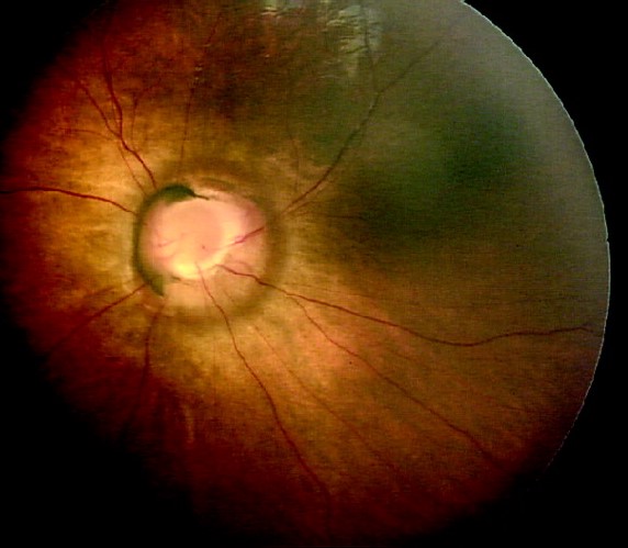
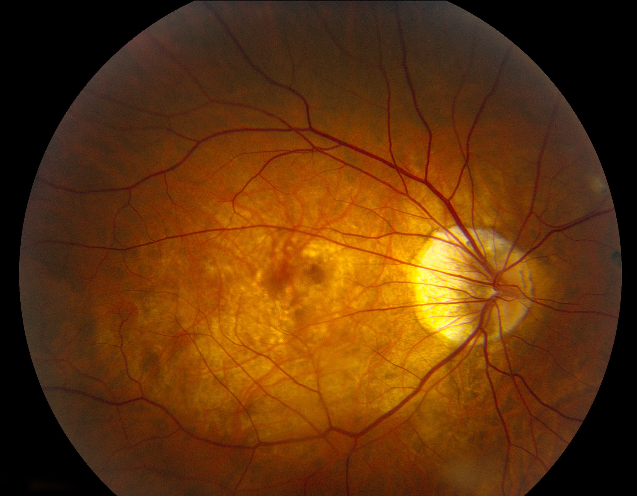
Unlike an optic disc pit or coloboma, which represent defects in the disc or nerve tissue itself, a peripapillary staphyloma is a defect of the sclera, an outpouching of the scleral wall around the optic nerve that creates a deep excavation in which the disc appears to sit at the bottom of a steep-walled pit.
The optic nerve tissue itself is usually relatively normal, though it can be displaced or distorted by the surrounding scleral anomaly, and the retinal vessels typically emerge from the disc and dip down into the excavation before spreading out onto the retina.
Clinical Features
- Usually unilateral, presenting from birth, though it may not be noticed until later if vision in the affected eye is reasonably preserved
- A deep, steep-walled excavation surrounding the optic disc, visible on fundus examination and often more dramatic in appearance than the degree of functional visual loss would suggest
- Visual acuity is variable, sometimes near normal, particularly when the fovea is spared
- Visual field defects, often an enlarged blind spot or other localized defect, but typically not the progressive, characteristic patterns of glaucomatous field loss
- No elevation of intraocular pressure, an important distinguishing feature from glaucoma
- Associated findings can include mild myopia in the affected eye
Fundus Explorer Pro
Photograph the retinal findings described here with the phone already in your pocket — 22 D optics and built-in illumination in one handheld unit.
From Choroida — the team behind this siteDistinguishing From Glaucoma
- Intraocular pressure is normal in isolated peripapillary staphyloma, whereas glaucomatous cupping is (with rare exceptions such as normal-tension glaucoma) associated with elevated or previously elevated pressure
- The excavation in a staphyloma has a distinctive, deep, steep-walled, often asymmetric funnel shape that differs from the smooth, progressively enlarging cup of glaucoma
- Staphyloma is present from birth and stable over time, while glaucomatous cupping progresses if untreated and stabilizes only with pressure control
- OCT and careful clinical photography, compared over time, show a static appearance in staphyloma versus progressive rim thinning in glaucoma
- Family and personal history of glaucoma risk factors supports that diagnosis, while an isolated, longstanding, unilateral deep excavation without other findings favors a congenital anomaly
Differential Diagnosis
- Optic disc coloboma, a related but distinct congenital anomaly that more often involves the disc and adjacent retina and choroid directly, sometimes with associated systemic findings (see coloboma)
- Morning glory disc anomaly, which has a distinctive funnel shape with a central glial tuft and radiating vessels, a different and usually more visually significant condition (see morning glory syndrome)
- Optic disc pit, a smaller, more focal excavation of the disc tissue itself rather than the surrounding sclera (see optic disc pit maculopathy)
- True glaucomatous cupping, distinguished as above
Evaluation
- Careful fundus photography and, when possible, comparison with any prior images to establish stability over time
- Intraocular pressure measurement, essential to exclude coexisting or superimposed glaucoma
- Visual field testing to characterize any deficit and to serve as a baseline for future comparison
- OCT of the optic nerve and retinal nerve fiber layer, interpreted cautiously given that normative databases are not designed for this anomalous disc architecture
Management
There is no treatment for the staphyloma itself, since it is a stable congenital anomaly rather than a progressive disease.
Management consists of correcting any refractive error, monitoring for amblyopia in children, and periodic follow-up to confirm stability, with prompt investigation of any new visual field change or reduction in acuity, since a coexisting or new problem should not be assumed to be part of the staphyloma without evidence of genuine progression.
Prognosis
Vision is typically stable over a patient’s lifetime once the diagnosis is established, and the main clinical importance of peripapillary staphyloma is being recognized correctly so that it is not mistaken for glaucoma, avoiding both unnecessary treatment and the anxiety of a presumed progressive diagnosis.


Document what you see
Two smartphone imaging tools built for everyday clinic use — one for the slit lamp, one for the fundus.
From Choroida — the team behind this siteReferences
- Cennamo G, Vecchio EC, de Crecchio G, Cennamo G. Evaluation of peripapillary staphyloma with optical coherence tomography. Retina. 2013;33:2099-2104.
- Bansal AS, Hsu J, Garg SJ, Sivalingam A, Vander JF. Optical coherence tomographic angiography of peripapillary staphyloma. JAMA Ophthalmol. 2016;134:e161831.
- Ceynowa DJ, Wickstrom R, Olsson M, et al. Optic disc anomalies in children: prevalence, ocular associations and neuroimaging findings. Acta Ophthalmol. 2019;97:582-589.
- Brodsky MC. Pediatric Neuro-Ophthalmology. 3rd ed. New York: Springer; 2016.
Test yourself
A few questions straight from this article.
-
A peripapillary staphyloma is a congenital defect of which structure?
Unlike an optic disc pit or coloboma, which are defects of disc or nerve tissue, a peripapillary staphyloma is an outpouching of the scleral wall around the optic nerve. -
Which finding most reliably separates isolated peripapillary staphyloma from glaucomatous cupping?
Intraocular pressure is normal in isolated peripapillary staphyloma, whereas glaucomatous cupping is associated with elevated or previously elevated pressure, rare exceptions aside. -
How do the retinal vessels behave at a peripapillary staphyloma on fundus examination?
Vessels typically emerge from the disc and dip down into the excavation before spreading out onto the surrounding retina. -
How does the excavation of a peripapillary staphyloma differ in shape from a glaucomatous cup?
The staphylomatous excavation is a distinctive deep, steep-walled, often asymmetric funnel, unlike the smooth, progressively enlarging cup of glaucoma. -
On serial optic nerve imaging, peripapillary staphyloma shows which behaviour?
Staphyloma is present from birth and stable, so OCT and serial photography show a static appearance, in contrast to the progressive rim thinning of glaucoma. -
Which refractive finding is associated with the eye affected by a peripapillary staphyloma?
Associated findings can include mild myopia in the affected eye. -
Which anomaly is a smaller, focal excavation of the disc tissue rather than the surrounding sclera?
An optic disc pit is a smaller, more focal excavation of the disc tissue itself, rather than of the sclera surrounding the nerve. -
Which features identify morning glory disc anomaly rather than peripapillary staphyloma?
Morning glory disc anomaly has a distinctive funnel shape with a central glial tuft and radiating vessels, and is usually more visually significant. -
Why must OCT nerve fibre layer values be read cautiously in peripapillary staphyloma?
OCT of the nerve and retinal nerve fibre layer is interpreted cautiously because normative databases are not designed for this anomalous disc architecture. -
What does management of a peripapillary staphyloma consist of?
There is no treatment for the anomaly itself: management is refractive correction, monitoring children for amblyopia, and periodic review to confirm stability.
