
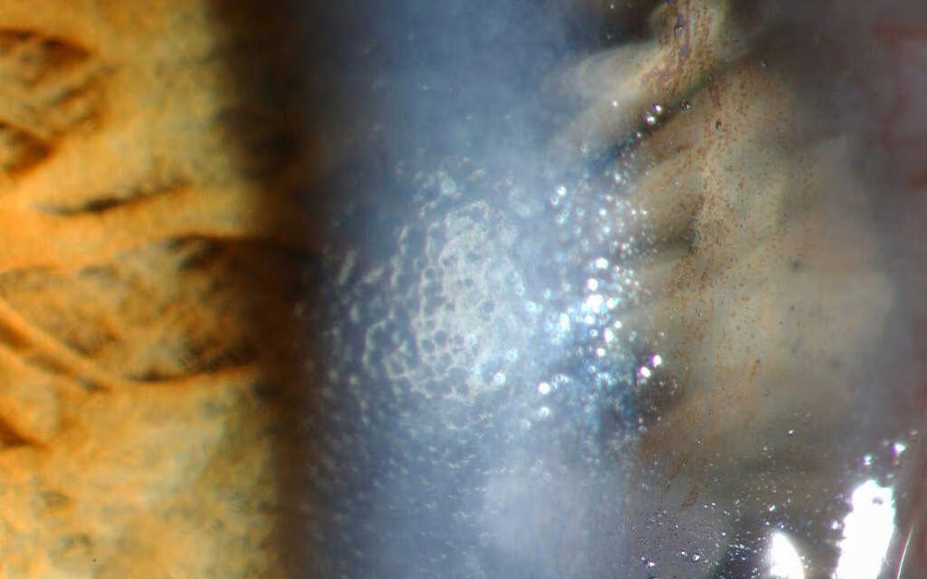
Fuchs endothelial corneal dystrophy is a bilateral, progressive degeneration of the corneal endothelium — the single layer of cells responsible for actively pumping fluid out of the cornea to keep it clear — and it is among the most common indications for corneal transplantation worldwide.
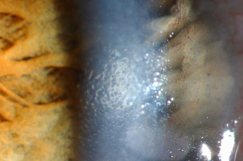
Its slow, decades-long course means many patients live with subclinical disease for years before vision is ever affected, which makes recognizing the earliest signs, well before symptoms develop, clinically useful for both counseling and surgical planning around any future intraocular surgery.
Pathogenesis
The disease is characterized by accelerated loss of corneal endothelial cells, well beyond the slow, expected age-related decline, combined with abnormal deposition of extracellular matrix material by the remaining dysfunctional cells, forming the excrescences on Descemet’s membrane known as guttae.
As endothelial cell density falls below the threshold needed to maintain adequate fluid pump function, the cornea begins to retain fluid, leading to progressive stromal and eventually epithelial edema.
A significant proportion of cases have an identifiable genetic component (mutations in genes including COL8A2 and, more commonly, a trinucleotide repeat expansion in TCF4), with a strong female predominance in most populations studied, typically an autosomal dominant inheritance pattern with variable penetrance in familial cases.
Clinical Staging
Early disease shows scattered central guttae without any visual symptoms, often found incidentally during a routine exam performed for another reason.
As guttae become more confluent and endothelial function declines further, patients begin to notice characteristic morning blurring that improves over the day.
Overnight, with the eyes closed, evaporation from the ocular surface is reduced, allowing more fluid to accumulate in the cornea, which then partially clears as the day progresses and evaporation resumes with the eyes open.
In advanced disease, this diurnal pattern is lost as corneal edema becomes constant, and epithelial bullae can form, causing pain in addition to visual blur when they rupture.
Tracking a patient through these stages — from asymptomatic guttae, through morning-predominant blur, to constant edema and eventual bullae — gives a useful, clinically intuitive framework for counseling patients about what to expect and for deciding when medical measures are likely to have reached their limit.
All-fit Slit-Lamp Adapter
Record and share exactly what you see at the slit lamp. One adapter fits any slit lamp or surgical microscope — and any smartphone.
From Choroida — the team behind this siteClinical Findings
- Guttae — dark, round excrescences on the corneal endothelium, best seen on specular reflection at the slit lamp, giving a “beaten metal” appearance in more advanced disease
- Stromal edema and thickening, measurable by pachymetry, which is a useful objective marker for tracking disease progression over time
- Epithelial edema and, in advanced cases, bullae — fluid-filled blisters on the corneal surface that can rupture and cause significant pain
- Characteristic diurnal variation in vision in moderate disease — worse on waking, improving over the day
- Reduced endothelial cell density and increased cell size/shape variability on specular microscopy, where available
Differential Diagnosis
- Posterior polymorphous corneal dystrophy — a different, generally more benign endothelial dystrophy with a distinct appearance and typically milder course
- Pseudophakic or aphakic bullous keratopathy — endothelial decompensation following cataract or other intraocular surgery, mechanistically distinct (surgical trauma rather than primary dystrophy) though the end-stage clinical picture can look similar
- Iridocorneal endothelial (ICE) syndrome — typically unilateral, with associated iris abnormalities, distinguishing it from the bilateral, symmetric pattern of Fuchs dystrophy
- Age-related cornea guttata without clinically significant dystrophy — very common as an incidental finding, representing one end of a spectrum that overlaps with true Fuchs dystrophy
Relevance to Cataract Surgery
Because cataract surgery itself causes some degree of endothelial cell loss even when performed without complication, a patient with pre-existing Fuchs dystrophy has less endothelial reserve to tolerate that additional insult, and can develop postoperative corneal decompensation even from routine, technically uncomplicated surgery.
Recognizing subclinical Fuchs dystrophy before cataract surgery — through careful slit-lamp exam for guttae and, where available, specular microscopy or pachymetry — allows for informed patient counseling and, in some cases, consideration of combined cataract and endothelial keratoplasty when the endothelial reserve is judged insufficient to tolerate cataract surgery alone, rather than discovering the problem only after an unexpectedly poor postoperative recovery.
Management
Early, asymptomatic disease requires no treatment beyond monitoring, with reassurance that most patients with mild, incidental guttae never progress to visually significant disease.
Symptomatic edema is initially managed with hypertonic saline drops or ointment, which draw fluid osmotically out of the cornea and can meaningfully improve morning symptoms in mild to moderate disease, along with a hair dryer held at arm’s length to gently dehydrate the corneal surface on waking, a simple measure some patients find helpful.
Endothelial keratoplasty — Descemet’s stripping automated endothelial keratoplasty (DSAEK) or, increasingly, Descemet membrane endothelial keratoplasty (DMEK) — has become the standard surgical treatment once medical measures no longer control symptoms, replacing only the diseased endothelial layer rather than the full-thickness cornea and offering faster visual recovery and better outcomes than traditional penetrating keratoplasty for this specific indication.
This shift away from full-thickness transplantation toward selective, layer-specific endothelial replacement represents one of the more significant advances in corneal surgery over recent decades, meaningfully changing the visual and recovery expectations that can be offered to patients with this common dystrophy.


Document what you see
Two smartphone imaging tools built for everyday clinic use — one for the slit lamp, one for the fundus.
From Choroida — the team behind this siteReferences
- Eghrari AO, Riazuddin SA, Gottsch JD. Fuchs corneal dystrophy. Progress in Molecular Biology and Translational Science.
- Zhang X, Igo RP Jr, Fondran J, et al. Association of smoking and other risk factors with Fuchs’ endothelial corneal dystrophy severity. Investigative Ophthalmology & Visual Science.
- Price MO, Price FW Jr. Endothelial keratoplasty — a review. Clinical and Experimental Ophthalmology.
- American Academy of Ophthalmology. Basic and Clinical Science Course, Section 8: External Disease and Cornea.
