
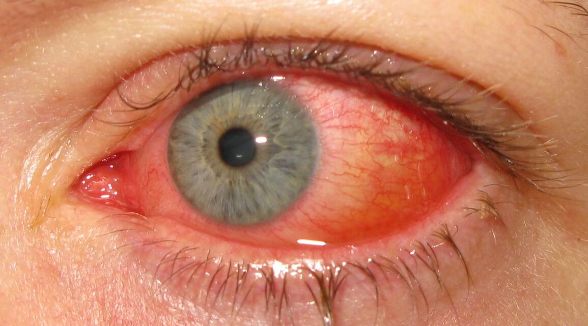
Few eye infections spread through a clinic waiting room as efficiently as this one — and few leave a mark on the cornea that lingers for months after the redness clears.

Epidemic keratoconjunctivitis (EKC) is a highly contagious viral conjunctivitis, almost always caused by adenovirus.
It earns the word “epidemic” honestly — outbreaks spread rapidly through households, schools, and eye clinics themselves when infection control lapses.
The acute redness resolves in most patients within one to two weeks, but a subset go on to develop corneal infiltrates that blur vision for weeks to months afterward.
There is no specific antiviral cure, which makes infection control and appropriate supportive care the two things that actually matter most.
Recognizing EKC promptly — and isolating it appropriately — protects both the patient’s cornea and everyone else in the waiting room.
What Is Epidemic Keratoconjunctivitis?
Epidemic keratoconjunctivitis (EKC) is an acute conjunctivitis, frequently with secondary corneal involvement, caused by specific adenovirus serotypes.
Its defining features include:
- Extremely high contagiousness, spread by direct contact, contaminated surfaces, and improperly disinfected clinical instruments
- A biphasic course — acute conjunctivitis followed, in a subset of patients, by delayed corneal subepithelial infiltrates
- No specific effective antiviral treatment, unlike some other ocular viral infections
- A well-documented history of causing clinic-based outbreaks when hand hygiene and instrument disinfection are inadequate
EKC is as much an infection-control problem as it is a clinical diagnosis — how it is managed in a waiting room matters almost as much as how it is treated in the patient.
Epidemiology
EKC is one of the most common causes of acute infectious conjunctivitis worldwide.
- Adenovirus serotypes 8, 19, and 37 are most classically associated with the keratoconjunctivitis (corneal-involving) form
- Outbreaks cluster in schools, workplaces, households, and healthcare settings, including ophthalmology clinics
- Transmission occurs readily via contaminated hands, towels, and ophthalmic equipment such as tonometer tips
- The virus can survive on surfaces for extended periods, contributing to its outbreak potential
A cluster of several patients with acute red eye presenting to the same clinic within days of each other should immediately raise suspicion for an EKC outbreak.
Pathophysiology
The disease unfolds in two distinct phases, which is why its course can seem to worsen even as the initial infection clears.
- Adenovirus directly infects the conjunctival epithelium, producing acute follicular conjunctivitis
- Viral replication and the resulting local inflammatory response drive the intense redness, chemosis, and watery discharge of the acute phase
- In a subset of patients, viral antigen deposited in the corneal subepithelial space triggers a delayed immune response
- This immune reaction produces subepithelial infiltrates that can appear one to two weeks after the acute conjunctivitis has already started improving
- These infiltrates represent an immune phenomenon rather than ongoing active viral replication, which is why they do not respond to antiviral therapy
Understanding that the corneal infiltrates are immune-mediated, not infectious, is exactly what explains why steroids — not antivirals — are the treatment when they become visually significant.
Risk Factors
Transmission Risk Factors
- Close contact with an infected individual, including household and school contacts
- Recent visit to an eye clinic, particularly where hand hygiene or instrument disinfection was inadequate
- Shared towels, pillows, or cosmetics with an infected person
Other Considerations
- No particular predisposition by age or sex — susceptibility is essentially universal on exposure
- Healthcare workers and eye care staff are at meaningfully elevated occupational risk during outbreaks
Because transmission is so efficient, the single most effective “risk factor modification” is strict hand hygiene and instrument disinfection around every red-eye patient, not just confirmed cases.
Clinical Presentation
Symptoms
- Acute redness, watery discharge, and foreign-body sensation, often starting in one eye and spreading to the other within days
- Preauricular lymph node tenderness or swelling, a helpful supporting clue
- Photophobia and blurred vision once corneal involvement develops
- A history of a recently affected close contact or a recent eye clinic visit
Examination Findings
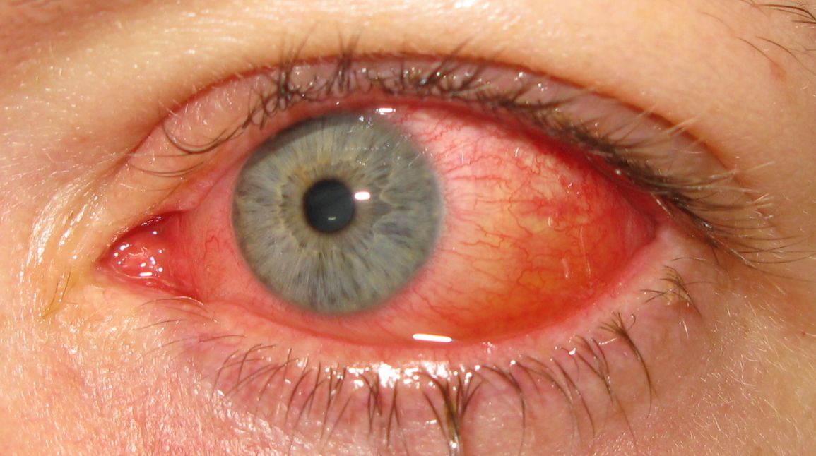
- Intense, diffuse bulbar conjunctival injection, often strikingly bright red
- Conjunctival chemosis and watery, non-purulent discharge
- Follicular reaction of the palpebral conjunctiva
- A tender, palpable preauricular lymph node on the affected side
- Fine, multiple corneal subepithelial infiltrates on slit-lamp exam, typically appearing one to two weeks into the illness
- Occasionally, a conjunctival pseudomembrane in more severe cases
The combination of preauricular adenopathy and follicular conjunctivitis is a classic, high-yield bedside pairing that should immediately suggest a viral rather than bacterial cause.
Diagnostic Evaluation
Clinical Examination
- Diagnosis is primarily clinical, based on the characteristic acute presentation and preauricular adenopathy
- Slit-lamp examination for subepithelial infiltrates, particularly in patients with persistent symptoms beyond the first week
Laboratory Testing
- Point-of-care adenovirus immunoassays can confirm the diagnosis rapidly where available
- PCR testing is more sensitive but generally reserved for outbreak investigation or diagnostically uncertain cases
Testing is rarely necessary for routine management — the clinical picture is usually distinctive enough on its own, and the main value of confirmation is for outbreak documentation.
Differential Diagnosis
Conditions that can be confused with EKC include:
- Bacterial conjunctivitis — purulent rather than watery discharge, and typically without significant preauricular adenopathy
- Allergic conjunctivitis — itching predominates, bilateral from onset, and lacks preauricular adenopathy
- Herpes simplex keratoconjunctivitis — dendritic corneal ulcers rather than subepithelial infiltrates, often unilateral
- Trachoma — follicular conjunctivitis but with a chronic, endemic-exposure pattern rather than an acute outbreak
Preauricular adenopathy combined with a watery, non-purulent discharge is the most useful bedside combination for separating EKC from its bacterial and allergic mimics.
Management
Acute Phase
- Supportive care — cold compresses, lubrication, and symptomatic relief — since no antiviral agent reliably shortens the acute illness
- Strict hygiene counseling: frequent hand washing, avoiding shared towels, and avoiding touching the eyes to limit spread to others
- Topical antibiotics are not indicated for the viral infection itself, though may be considered if secondary bacterial infection is suspected
Subepithelial Infiltrates
- Observation alone for infiltrates that are not visually significant, since most fade gradually over weeks to months
- Topical corticosteroids for infiltrates causing significant visual symptoms, since the infiltrates are immune-mediated rather than infectious
- Steroids should be tapered carefully, since infiltrates can recur or rebound if stopped too quickly
Infection Control
- Rigorous hand hygiene and disinfection of any instrument that contacts the eye between patients
- Isolating known or suspected cases within the clinic flow where practical
- Advising affected patients to stay home from school or work while actively contagious
Infection control measures are not an afterthought in EKC — they are as central to management as anything prescribed for the individual patient.
Prognosis
Prognosis is generally good, though the corneal phase can be prolonged.
- Acute conjunctivitis typically resolves within one to two weeks
- Subepithelial infiltrates can persist for weeks to several months, and occasionally longer, before fully clearing
- Permanent visual impairment is uncommon but can occur with dense, persistent central infiltrates
Most of the lingering concern in EKC comes from the corneal phase, not the acute conjunctivitis — setting expectations about that delayed course helps patients tolerate a slow recovery.
Would you like to document anterior segment findings with your smartphone?
Smartphone slit-lamp photography makes it easy to document conjunctival injection and corneal subepithelial infiltrates in epidemic keratoconjunctivitis and track them over the course of recovery using a simple slit-lamp adaptor.
SLIT-LAMP SMARTPHONE PHOTOGRAPHY
References
- Jhanji V, Chan TC, Li EY, et al. Adenoviral keratoconjunctivitis. Survey of Ophthalmology. 2015.
- American Academy of Ophthalmology. Conjunctivitis Preferred Practice Pattern. 2024.
- Meyer-Rusenberg B, Loderstadt U, Richard G, et al. Epidemic keratoconjunctivitis: the current situation and recommendations for prevention and treatment. Deutsches Arzteblatt International. 2011.
- Epidemic Keratoconjunctivitis. EyeWiki, American Academy of Ophthalmology.
- Kaufman HE. Adenovirus advances: new diagnostic and therapeutic options. Current Opinion in Ophthalmology. 2011.

