
A single fundus photograph can already tell you which patients are at risk of a second, far more dangerous diagnosis weeks later.
Central retinal vein occlusion (CRVO) begins as blocked venous drainage from the entire retina, not just a sector of it.
That distinction from its more common cousin, branch retinal vein occlusion, is what makes CRVO both more dramatic at presentation and more dangerous afterward.
Roughly one in five cases is the ischemic subtype, and ischemic eyes carry a real risk of progressing to neovascular glaucoma.
Getting the ischemic-versus-non-ischemic classification right at diagnosis — and monitoring accordingly — is what actually prevents that second, more devastating complication.
Understanding the fundus pattern, the diagnostic workup, and the anti-VEGF era of treatment is essential for managing this disease safely.
What Is Central Retinal Vein Occlusion?
Central retinal vein occlusion (CRVO) is obstruction of the central retinal vein at or near the optic nerve head, blocking venous outflow from the entire retina.
CRVO is classified into two clinically distinct forms:
- Non-ischemic CRVO — the more common form, with better visual prognosis and lower risk of neovascular complications
- Ischemic CRVO — roughly one in five cases, with extensive capillary nonperfusion, worse visual outcomes, and a substantial risk of neovascular glaucoma
Because the entire retina drains through this single vein, occlusion here produces far more widespread hemorrhage than a branch occlusion ever does.
Epidemiology
CRVO is less common than branch retinal vein occlusion but carries a meaningfully higher complication rate.
- It occurs roughly a quarter as often as BRVO in most population-based studies
- Approximately 20% of CRVO cases are the ischemic subtype at presentation
- Prevalence rises with age, and shares many of the same cardiovascular risk factors as BRVO
- Bilateral involvement, though uncommon, occurs more often than in the general population, particularly with underlying systemic vascular disease
Because ischemic and non-ischemic CRVO follow such different clinical courses, epidemiologic figures for “CRVO” as a whole can be misleading without separating the two subtypes.
Fundus Explorer Pro
Photograph the retinal findings described here with the phone already in your pocket — 22 D optics and built-in illumination in one handheld unit.
From Choroida — the team behind this sitePathophysiology
The disease mechanism mirrors BRVO but at the level of the entire central retinal vein rather than a single branch.
- Thrombosis develops at or near the lamina cribrosa, where the central retinal vein is most vulnerable to compression and turbulent flow
- Elevated venous pressure backs up throughout the entire retinal venous system
- Widespread capillary leakage produces hemorrhage and macular edema across all four quadrants
- In more severe occlusion, capillary nonperfusion produces retinal ischemia, which drives the release of vascular endothelial growth factor (VEGF)
- VEGF release can trigger neovascularization of the iris and angle, the mechanism behind neovascular glaucoma
The degree of capillary nonperfusion is the single factor that most determines whether a given eye will follow the relatively benign non-ischemic course or the higher-risk ischemic one.
Risk Factors
Systemic Risk Factors
- Systemic hypertension
- Diabetes mellitus
- Hyperlipidemia
- Hypercoagulable states, more relevant in younger patients with bilateral or atypical presentations
Ocular Risk Factors
- Elevated intraocular pressure or glaucoma
- Older age
- Open-angle glaucoma specifically has been associated with increased CRVO risk in several studies
The overlap with cardiovascular risk factors means every new CRVO diagnosis is also an opportunity to check that a patient’s systemic vascular risk is being properly managed.
Clinical Presentation
Symptoms
- Sudden, painless vision loss, typically more pronounced than in BRVO given the diffuse retinal involvement
- Vision loss can range from mild blurring in non-ischemic disease to severe, profound loss in ischemic CRVO
- Eye pain, if present, should raise concern for secondary neovascular glaucoma rather than the occlusion itself
Examination Findings
- Diffuse flame-shaped and blot hemorrhages scattered across all four quadrants — the classic “blood and thunder” fundus
- Dilated, tortuous retinal veins throughout the fundus
- Optic disc edema and macular edema in many eyes
- Cotton-wool spots, more numerous and extensive in ischemic disease
- A relative afferent pupillary defect, particularly in ischemic CRVO, reflecting the degree of retinal dysfunction
Unlike BRVO’s sector-confined pattern, CRVO hemorrhage genuinely involves all four quadrants — that distribution alone is usually enough to make the diagnosis at a glance.
Diagnostic Evaluation
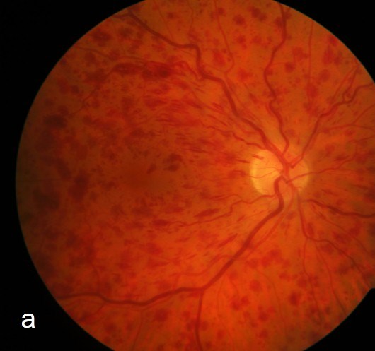
Fundus Examination and Photography
- Dilated exam or wide-field photography to document the extent of hemorrhage and disc involvement
- Assessment of relative afferent pupillary defect, which correlates with the degree of ischemia
Optical Coherence Tomography
- Quantifies macular edema and helps guide anti-VEGF treatment decisions
- Serial OCT tracks response to treatment over time
Fluorescein Angiography
- Defines the extent of capillary nonperfusion, the key determinant of ischemic versus non-ischemic classification
- Ten or more disc areas of nonperfusion is a widely used threshold for defining ischemic CRVO
Ongoing Monitoring for Neovascularization
- Regular gonioscopy and iris examination for early neovascularization, particularly in ischemic or indeterminate cases
- Close follow-up during the first six months, when the risk of neovascular glaucoma is highest
The angiographic extent of nonperfusion, more than the hemorrhage seen on the fundus photo alone, is what actually determines an eye’s risk of neovascular glaucoma.
Differential Diagnosis
Conditions that can resemble or coexist with CRVO include:
- Branch retinal vein occlusion — hemorrhage confined to a single sector rather than all four quadrants
- Ocular ischemic syndrome — mid-peripheral hemorrhages with additional signs of carotid insufficiency, including anterior segment ischemia
- Diabetic retinopathy — typically more symmetric between the two eyes and progresses over a longer timeframe
- Hypertensive retinopathy — arteriolar changes and hemorrhages without the venous dilation and tortuosity of CRVO
- Papilledema — disc edema without the diffuse four-quadrant hemorrhagic pattern
The combination of diffuse four-quadrant hemorrhage, venous dilation, and disc edema is distinctive enough that CRVO is rarely confused with these mimics once the full fundus is examined.
Management
Macular Edema
- Intravitreal anti-VEGF therapy (ranibizumab, aflibercept, or bevacizumab) is first-line treatment for vision-threatening macular edema
- Intravitreal corticosteroid implants are an alternative, particularly in pseudophakic eyes or when anti-VEGF response is inadequate
- Regular re-treatment is typically needed, since edema commonly recurs when therapy is paused
Neovascular Complications
- Panretinal photocoagulation is indicated once iris, angle, or retinal neovascularization develops
- Anti-VEGF therapy reduces the incidence of neovascular complications and can be used adjunctively, though it does not eliminate the risk in ischemic eyes
- Prompt treatment of neovascular glaucoma, including IOP-lowering therapy and, when needed, glaucoma surgery, is critical once it develops
Monitoring and Follow-Up
- Frequent follow-up during the first six months after diagnosis, the period of highest risk for neovascular glaucoma in ischemic eyes
- Ongoing systemic risk factor management in coordination with the patient’s primary care physician
The treatment plan for CRVO is really two parallel tracks — managing macular edema for vision, and actively surveilling for neovascularization to prevent a second, separate disaster.
Prognosis
Visual outcome depends heavily on ischemic status and how promptly neovascular complications are caught.
- Non-ischemic CRVO carries a comparatively favorable visual prognosis, especially with anti-VEGF treatment of macular edema
- Ischemic CRVO carries a substantially worse visual prognosis and a real risk of neovascular glaucoma, historically reported in roughly a quarter to more than half of untreated ischemic eyes
- Anti-VEGF therapy has meaningfully reduced, though not eliminated, the incidence of neovascular glaucoma in treated eyes
The single biggest driver of long-term outcome is not the initial hemorrhage but whether neovascular glaucoma is caught and treated before it causes irreversible damage.


Document what you see
Two smartphone imaging tools built for everyday clinic use — one for the slit lamp, one for the fundus.
From Choroida — the team behind this siteReferences
- Central Vein Occlusion Study Group. Baseline and early natural history report. Archives of Ophthalmology. 1993.
- Brown DM, Campochiaro PA, Singh RP, et al. Ranibizumab for macular edema following central retinal vein occlusion: the CRUISE study. Ophthalmology. 2010.
- Rogers S, McIntosh RL, Cheung N, et al. The prevalence of retinal vein occlusion: pooled data from population studies. Ophthalmology. 2010.
- The progress of assessment methods and treatments of neovascular glaucoma secondary to central retinal vein occlusion. Frontiers in Medicine. 2024.
- Central Retinal Vein Occlusion. EyeWiki, American Academy of Ophthalmology.
Test yourself
A few questions straight from this article.
-
Approximately what proportion of central retinal vein occlusion cases are the ischemic subtype at presentation?
Roughly one in five CRVO cases — about 20% — are the ischemic subtype at presentation, and those eyes carry a real risk of progressing to neovascular glaucoma. -
Where does the thrombus in central retinal vein occlusion typically form?
Thrombosis develops at or near the lamina cribrosa, where the central retinal vein is most vulnerable to compression and turbulent flow. -
Which fundus pattern distinguishes CRVO from branch retinal vein occlusion?
CRVO produces diffuse flame-shaped and blot hemorrhages across all four quadrants — the classic "blood and thunder" fundus. BRVO hemorrhage stays confined to one sector. -
On fluorescein angiography, what extent of capillary nonperfusion is the widely used threshold for defining ischemic CRVO?
Ten or more disc areas of nonperfusion is a widely used threshold for classifying a CRVO as ischemic. -
A patient with known CRVO returns complaining of eye pain. What should this raise concern for?
Pain is not a feature of the occlusion itself. Its appearance should raise concern for secondary neovascular glaucoma. -
What is first-line treatment for vision-threatening macular edema in CRVO?
Intravitreal anti-VEGF therapy (ranibizumab, aflibercept or bevacizumab) is first line. Corticosteroid implants are an alternative, particularly in pseudophakic eyes or when the anti-VEGF response is inadequate. -
During which period after diagnosis is the risk of neovascular glaucoma highest?
The first six months after diagnosis carry the highest risk of neovascular glaucoma, which is why close follow-up is concentrated in that window. -
Which single factor most determines whether a CRVO eye follows the non-ischemic or the ischemic course?
The angiographic extent of nonperfusion, more than the hemorrhage visible on the fundus photograph, is what actually determines an eye's risk of neovascular glaucoma. -
Which examination finding correlates with the degree of ischemia in CRVO?
A relative afferent pupillary defect, particularly in ischemic CRVO, reflects the degree of retinal dysfunction, and assessing it is part of the standard workup.

Writes and reviews the clinical articles on Educate. Every post is checked against peer-reviewed sources before it goes live.


