
A single crossing point on the retina can decide whether a patient keeps or loses central vision.
Branch retinal vein occlusion (BRVO) begins at exactly that point — where a retinal artery and vein share a common sheath.
It is the second most common retinal vascular disease after diabetic retinopathy, and the most common of the retinal vein occlusions.
The presentation can range from an incidental finding on routine fundus exam to sudden, painless vision loss.
What happens next depends heavily on whether the occlusion involves the macula and whether the retina becomes ischemic.
Recognizing the pattern early, grading its severity correctly, and choosing the right treatment window can be the difference between a full visual recovery and permanent central vision loss.
What Is Branch Retinal Vein Occlusion?
Branch retinal vein occlusion (BRVO) is an obstruction of one of the branch retinal veins, typically at an arteriovenous crossing, that impairs venous drainage from the corresponding sector of retina.
BRVO is generally classified as:
- Major BRVO — occlusion of a first-order branch vein, affecting a large retinal quadrant
- Macular BRVO — occlusion of a smaller venule draining only the macula
- Ischemic BRVO — significant capillary nonperfusion on angiography, with higher risk of neovascular complications
- Non-ischemic BRVO — the more common and generally better-prognosis form
The superotemporal quadrant is affected far more often than any other, simply because it contains the greatest number of arteriovenous crossings.
Epidemiology
BRVO is common enough that most retina clinics see new cases regularly.
- BRVO affects an estimated 20 million or more people worldwide
- It is roughly four times more prevalent than central retinal vein occlusion (CRVO)
- Prevalence rises steadily with age, with most patients presenting after age 50
- Hypertension, diabetes mellitus, and dyslipidemia are the most consistently reported systemic associations
As the population ages and cardiovascular risk factors remain widespread, BRVO will continue to be encountered frequently in everyday retina practice.
Fundus Explorer Pro
Photograph the retinal findings described here with the phone already in your pocket — 22 D optics and built-in illumination in one handheld unit.
From Choroida — the team behind this sitePathophysiology
Retinal arteries and veins share a common adventitial sheath wherever they cross.
The sequence that leads to occlusion typically involves:
- A rigid, arteriosclerotic artery compressing the adjacent vein at the shared crossing point
- Turbulent blood flow and endothelial damage at the site of compression
- Secondary thrombus formation within the compressed vein
- Elevated venous pressure and stasis in the drainage territory beyond the occlusion
- Vascular leakage producing hemorrhage and macular edema, and — if severe — capillary nonperfusion and retinal ischemia
This is why BRVO is fundamentally a disease of vascular crossings, and why systemic conditions that stiffen arterial walls raise the risk directly.
Risk Factors
Systemic Risk Factors
- Systemic hypertension — the single most consistently reported risk factor
- Diabetes mellitus
- Dyslipidemia
- Elevated body mass index
- Hypercoagulable states, including antiphospholipid syndrome, in selected younger patients
Ocular Risk Factors
- Older age
- A history of glaucoma or elevated intraocular pressure
- Arteriovenous crossing anatomy where the artery lies anterior to the vein
- Oral contraceptive use has been reported as a contributing factor in some series
Controlling systemic vascular risk factors is as much a part of BRVO management as treating the eye itself.
Clinical Presentation
Symptoms
Presentation varies widely depending on whether the macula is involved.
- Sudden, painless, sector-shaped visual field loss when a major branch is occluded
- Blurred or distorted central vision when the macula is involved by edema or ischemia
- No symptoms at all when the occlusion is peripheral and spares the macula, sometimes found incidentally
Examination Findings
The fundus appearance is often distinctive enough to diagnose on sight:
- Flame-shaped and blot hemorrhages confined to the drainage territory of the affected vein
- Dilated, tortuous venous segment distal to the occlusion
- Cotton-wool spots in more ischemic cases
- Retinal edema and hard exudates when the macula is involved
- A wedge- or sector-shaped distribution that respects the horizontal raphe — a hallmark that separates BRVO from CRVO
That sector-confined pattern, sparing the retina outside the affected vein’s drainage territory, is the single most useful sign at the slit lamp or indirect ophthalmoscope.
Diagnostic Evaluation
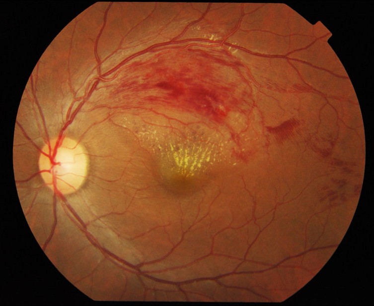
Fundus Examination and Photography
- Dilated fundus exam or wide-field photography to document the extent and location of hemorrhage
- Comparison with the fellow eye and serial photographs to track resolution or progression
Optical Coherence Tomography
- Quantifies macular edema and guides the decision to treat
- Detects intraretinal cystoid spaces, subretinal fluid, and ellipsoid zone disruption that correlates with visual prognosis
Fluorescein Angiography
- Defines the extent of capillary nonperfusion and distinguishes ischemic from non-ischemic BRVO
- Five or more disc areas of nonperfusion is the threshold traditionally used to define an ischemic, higher-risk occlusion
- Identifies areas of leakage and early neovascularization not always visible clinically
Systemic Workup
- Blood pressure measurement and basic metabolic and lipid panels in essentially all patients
- Additional hypercoagulability or inflammatory workup in patients under 50, those with bilateral disease, or an atypical systemic history
Angiography and OCT together determine both the treatment plan and the surveillance schedule for a given patient.
Differential Diagnosis
Conditions that can resemble or coexist with BRVO include:
- Central retinal vein occlusion — hemorrhage in all four quadrants rather than a single sector
- Hemi-retinal vein occlusion — occlusion at the disc affecting a full hemisphere
- Diabetic retinopathy — hemorrhages are typically more diffuse and not confined to a single vascular territory
- Hypertensive retinopathy — arteriolar changes without a sector-confined hemorrhagic pattern
- Ocular ischemic syndrome — mid-peripheral hemorrhages with retinal and often anterior segment ischemic signs
- Radiation retinopathy — relevant history of ocular or orbital radiotherapy
The sector-confined, raphe-respecting distribution of hemorrhage is usually enough to separate BRVO from these mimics on its own.
Management
Macular Edema
- Intravitreal anti-VEGF therapy (ranibizumab, aflibercept, or bevacizumab) is first-line for vision-threatening macular edema
- Intravitreal corticosteroid implants are a reasonable option, particularly in pseudophakic eyes or when anti-VEGF response is inadequate
- Macular grid laser photocoagulation, established by the Branch Vein Occlusion Study, remains a treatment option, though anti-VEGF therapy has largely become first-line
Neovascular Complications
- Sector scatter (panretinal) photocoagulation is indicated once retinal or disc neovascularization develops, or prophylactically in eyes with extensive nonperfusion under close follow-up
- Anti-VEGF therapy can also regress neovascularization, though laser remains the more durable long-term treatment
- Vitreous hemorrhage or tractional retinal detachment from neovascularization may require vitrectomy
Observation
- Peripheral BRVO without macular involvement and without significant ischemia can often be observed with periodic follow-up
- Underlying systemic risk factors should still be addressed even when the eye itself does not require treatment
Treatment decisions hinge on two questions: is the macula involved, and is the retina ischemic — both determine urgency and modality.
Prognosis
Visual outcome depends largely on:
- Whether the macula is involved by edema or ischemia
- Ischemic versus non-ischemic status at presentation
- Timeliness and response to anti-VEGF or steroid treatment
- Development of secondary complications such as neovascular glaucoma or vitreous hemorrhage
Many eyes with non-ischemic BRVO recover good vision with treatment of macular edema, while ischemic BRVO carries a higher risk of neovascular complications and a less favorable long-term outcome.


Document what you see
Two smartphone imaging tools built for everyday clinic use — one for the slit lamp, one for the fundus.
From Choroida — the team behind this siteReferences
- Rogers S, McIntosh RL, Cheung N, et al. The prevalence of retinal vein occlusion: pooled data from population studies from the United States, Europe, Asia, and Australia. Ophthalmology. 2010.
- Branch Vein Occlusion Study Group. Argon laser photocoagulation for macular edema in branch vein occlusion. American Journal of Ophthalmology. 1984.
- Campochiaro PA, Heier JS, Feiner L, et al. Ranibizumab for macular edema following branch retinal vein occlusion: the BRAVO study. Ophthalmology. 2010.
- Ho M, Liu DT, Lam DS, Jonas JB. Retinal vein occlusions, from basics to the latest treatment. Retina. 2016.
- The Diagnosis and Treatment of Branch Retinal Vein Occlusions: An Update. Journal of Clinical Medicine. 2025.
- EyeWiki (American Academy of Ophthalmology). Branch Retinal Vein Occlusion.
Test yourself
A few questions straight from this article.
-
Branch retinal vein occlusion typically obstructs a retinal vein at which site?
BRVO is fundamentally a disease of vascular crossings: artery and vein share a common adventitial sheath wherever they cross, and the occlusion begins at that point. -
Which retinal quadrant is affected most often in branch retinal vein occlusion?
The superotemporal quadrant is involved far more often than any other, simply because it contains the greatest number of arteriovenous crossings. -
How does the prevalence of branch retinal vein occlusion compare with central retinal vein occlusion?
BRVO is roughly four times more prevalent than CRVO and is the most common of the retinal vein occlusions, affecting an estimated 20 million or more people worldwide. -
What initiates the venous obstruction in branch retinal vein occlusion?
A stiff arteriosclerotic artery compresses the adjacent vein at the shared crossing, causing turbulent flow, endothelial damage and secondary thrombus formation. -
Which systemic condition is the most consistently reported risk factor for branch retinal vein occlusion?
Hypertension is the single most consistently reported risk factor, alongside diabetes mellitus and dyslipidemia as the other common systemic associations. -
Which fundus pattern is the hallmark of branch retinal vein occlusion?
Hemorrhages stay confined to the drainage territory of the occluded vein in a wedge- or sector-shaped distribution that respects the horizontal raphe, which separates BRVO from CRVO. -
What extent of capillary nonperfusion on angiography traditionally defines ischemic branch retinal vein occlusion?
Five or more disc areas of nonperfusion is the traditional threshold for an ischemic, higher-risk occlusion, which carries a greater chance of neovascular complications. -
What is first-line treatment for vision-threatening macular edema in branch retinal vein occlusion?
Intravitreal anti-VEGF agents such as ranibizumab, aflibercept or bevacizumab are first-line; macular grid laser remains an option but anti-VEGF has largely replaced it. -
Which treatment is indicated once retinal or disc neovascularization develops in branch retinal vein occlusion?
Sector scatter (panretinal) photocoagulation is indicated once neovascularization appears; anti-VEGF can also regress it, but laser is the more durable long-term treatment. -
Which feature predicts a less favourable long-term outcome in branch retinal vein occlusion?
Ischemic BRVO carries a higher risk of neovascular complications and a less favourable outcome, while many non-ischemic eyes recover good vision once macular edema is treated.

Writes and reviews the clinical articles on Educate. Every post is checked against peer-reviewed sources before it goes live.


