A painful red eye with a cloudy cornea and a mid-dilated pupil is an emergency until proven otherwise.
Few conditions in ophthalmology reward speed as directly as this one. Pressure that stays high for hours strangles the optic nerve, and vision lost to a missed attack rarely comes back.
Acute angle-closure glaucoma (AACG) is the classic sight-threatening emergency of the anterior segment — and one that non-ophthalmologists frequently mistake for conjunctivitis or migraine.
What Is Acute Angle-Closure Glaucoma?
AACG occurs when the peripheral iris suddenly blocks the trabecular meshwork, cutting off aqueous drainage. Intraocular pressure rises rapidly — often to 40–80 mmHg — causing pain, corneal edema, and, if unrelieved, optic nerve damage within hours.
The usual mechanism is pupillary block: aqueous cannot pass from the posterior to the anterior chamber through the pupil, so it pushes the iris forward and seals the angle. Understanding that the block is at the pupil is exactly why an iridotomy — a hole that bypasses it — is the definitive cure.
Who Is at Risk
The anatomy of the eye largely determines the risk.
- Hyperopic (long-sighted) eyes with shallow anterior chambers and short axial length.
- Older age, as the lens thickens and crowds the angle.
- Female sex and East Asian ethnicity.
- A narrow angle in the fellow eye — the single best predictor.
- Dim-light triggers and pupil-dilating drugs (anticholinergics, sympathomimetics, some antidepressants and decongestants) that leave the pupil mid-dilated.
Asking about a recent new medication or a first attack in a darkened cinema often uncovers the trigger.
All-fit Slit-Lamp Adapter
Record and share exactly what you see at the slit lamp. One adapter fits any slit lamp or surgical microscope — and any smartphone.
From Choroida — the team behind this siteClinical Presentation
The picture is dramatic when complete, but partial attacks mislead.
- Sudden, severe unilateral eye pain and blurred vision.
- Haloes around lights from corneal edema.
- Headache, nausea, and vomiting — so prominent that patients are sometimes worked up for an abdominal or neurological cause.
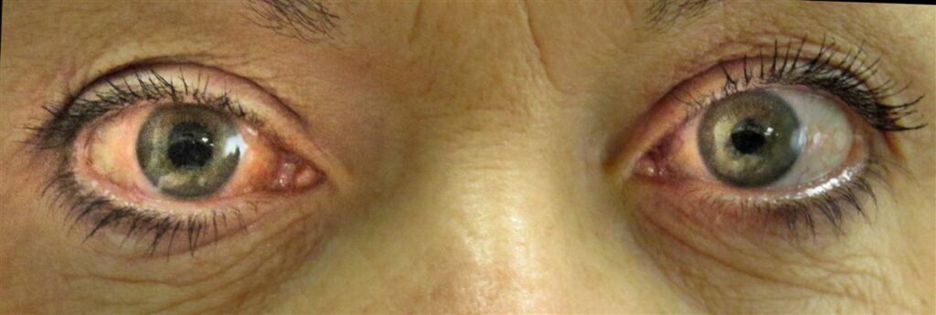
- A red, congested eye with a steamy cornea and a fixed, mid-dilated, vertically oval pupil.
- A rock-hard globe on gentle palpation and markedly raised intraocular pressure.
The combination of a mid-dilated non-reactive pupil with a hazy cornea in a painful red eye is the pattern that should stop you — conjunctivitis does neither.
Why It Is So Often Missed
Two traps recur. Systemic symptoms — vomiting and headache — can dominate, sending the patient to a medical or neurology service while the eye goes untreated.
The other trap is intermittent, self-terminating subacute attacks: brief evening episodes of blurred vision and haloes that resolve by morning. These “warning” attacks are easily dismissed, yet they identify an eye poised to close completely.
Taking any painful red eye with visual symptoms to the slit lamp and tonometer, rather than treating it empirically as infective, is what prevents the miss.
Diagnostic Evaluation
- Tonometry confirms the grossly elevated intraocular pressure.
- Slit-lamp examination shows corneal edema, a shallow anterior chamber, and a mid-dilated pupil.
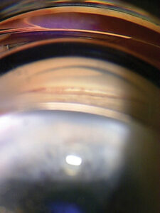
- Gonioscopy — ideally of the fellow eye — confirms an occludable angle and guides prophylaxis.
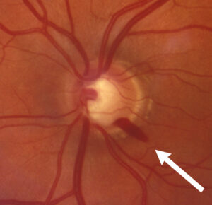
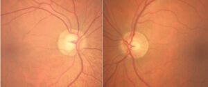
- Optic disc assessment once the cornea clears, to gauge any glaucomatous damage.
Examining and treating the fellow eye is not optional — it is anatomically primed for the same attack.
Differential Diagnosis
- Acute anterior uveitis — painful and red, but with a small or normal pupil and normal-to-low pressure.
- Infective conjunctivitis or keratitis — red but with a reactive pupil and normal pressure.
- Neovascular or other secondary glaucomas presenting with high pressure.
- Acute hydrops or scleritis as painful red-eye mimics.
Pupil size and intraocular pressure are the two bedside findings that most quickly separate AACG from its mimics.
Management
Treatment runs in two phases: break the attack medically, then cure it definitively.
- Immediate pressure-lowering with topical agents (beta-blocker, alpha-agonist, and a topical carbonic anhydrase inhibitor) plus systemic acetazolamide, and hyperosmotic agents when needed.
- Topical pilocarpine to constrict the pupil and pull the iris out of the angle — often more effective once the pressure begins to fall and the iris sphincter recovers.
- Supportive care for pain and vomiting, and prompt referral.
- Laser peripheral iridotomy is the definitive treatment, creating a channel that relieves pupillary block — performed in the affected eye once the cornea clears and prophylactically in the fellow eye.
- Lens extraction is increasingly used, since removing the thickened lens deepens the angle and can be definitive.
The two-step logic — medicine buys time, iridotomy (or lens surgery) fixes the anatomy — is the core of getting AACG right.
Prognosis
Vision usually recovers well when pressure is broken within hours. Delay is what converts a treatable emergency into permanent optic nerve damage, chronic angle-closure from peripheral anterior synechiae, or a fixed dilated pupil and glaukomflecken from the attack itself.
Because the fellow eye carries a high lifetime risk, prophylactic treatment turns a one-eye emergency into a fully prevented second event.
Would you like to document an acute red eye like angle-closure glaucoma with your smartphone?


Document what you see
Two smartphone imaging tools built for everyday clinic use — one for the slit lamp, one for the fundus.
From Choroida — the team behind this siteReferences
- Weinreb RN, Aung T, Medeiros FA. The pathophysiology and treatment of glaucoma: a review. JAMA. 2014;311(18):1901-1911.
- He M, Jiang Y, Huang S, et al. Laser peripheral iridotomy for the prevention of angle closure: a randomised controlled trial. The Lancet. 2019;393(10181):1609-1618.
- Azuara-Blanco A, Burr J, Ramsay C, et al. Effectiveness of early lens extraction for the treatment of primary angle-closure glaucoma (EAGLE): a randomised controlled trial. The Lancet. 2016;388(10052):1389-1397.
- European Glaucoma Society. Terminology and Guidelines for Glaucoma, 5th Edition. British Journal of Ophthalmology. 2021;105(Suppl 1):1-169.
Test yourself
A few questions straight from this article.
-
What is the usual mechanism of acute angle-closure glaucoma?
Aqueous cannot pass through the pupil from the posterior to the anterior chamber, so it bows the iris forward and seals the angle; that pupillary block is why an iridotomy is curative. -
To what range does intraocular pressure typically rise in an acute angle-closure attack?
Pressure rises rapidly, often to 40-80 mmHg, producing pain and corneal oedema and threatening the optic nerve within hours if unrelieved. -
What is the single best predictor of risk for acute angle-closure glaucoma?
A narrow angle in the fellow eye is the single best predictor of risk, which is why gonioscopy of that eye guides prophylactic treatment. -
Which anatomical profile predisposes an eye to acute angle-closure glaucoma?
Hyperopic eyes with shallow anterior chambers and short axial length are anatomically predisposed, and the risk rises further as the lens thickens with age. -
Which pupil finding characterises an acute angle-closure attack?
The fixed, mid-dilated, vertically oval pupil combined with a steamy cornea in a painful red eye is the pattern that separates angle closure from conjunctivitis. -
Which history suggests an eye poised for a complete acute angle-closure attack?
Intermittent, self-terminating subacute attacks cause brief evening blurring and haloes that resolve overnight; they are easily dismissed but identify an eye about to close completely. -
Which feature separates acute anterior uveitis from acute angle-closure glaucoma?
Acute anterior uveitis is painful and red but shows a small or normal pupil and normal-to-low intraocular pressure, whereas angle closure gives a mid-dilated pupil and very high pressure. -
Which regimen is used to break an acute angle-closure attack medically?
A topical beta-blocker, alpha-agonist and carbonic anhydrase inhibitor with systemic acetazolamide lower pressure immediately, with pilocarpine added to pull the iris out of the angle as pressure falls. -
What is the definitive treatment for acute angle-closure glaucoma?
Laser peripheral iridotomy creates a channel that relieves pupillary block; it is performed in the affected eye once the cornea clears and prophylactically in the fellow eye. -
Which lasting change can follow a delayed or prolonged angle-closure attack?
Delay converts a treatable emergency into permanent optic nerve damage, chronic angle closure from peripheral anterior synechiae, or a fixed dilated pupil with glaukomflecken.

Writes and reviews the clinical articles on Educate. Every post is checked against peer-reviewed sources before it goes live.