
Foster Kennedy Syndrome is a tale of two optic discs that never match.

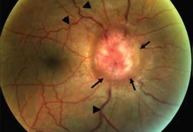
One nerve is pale and atrophic from long-standing compression, while the other is swollen from the raised pressure the same mass has caused.
The asymmetry is the whole point, and it can be the first sign of a large intracranial tumour.
For clinicians, recognizing Foster Kennedy Syndrome matters because the classic form signals a mass that needs imaging, while its common mimic needs no scan at all.
Comparing the two optic nerves side by side is what starts the reasoning.
What Is Foster Kennedy Syndrome?
Foster Kennedy Syndrome is the combination of optic atrophy in one eye and papilledema in the other, caused by a compressive intracranial mass.
Its defining features are:
- Optic atrophy in the eye on the side of the lesion, from direct compression of that optic nerve
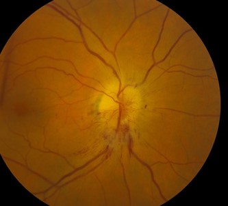
- Papilledema in the fellow eye, from the raised intracranial pressure the mass produces
- Central scotoma and reduced vision in the atrophic eye
- Frequently anosmia, when an olfactory groove or frontal lesion is responsible
The classic culprit is a frontal lobe or olfactory groove meningioma pressing on one nerve while raising pressure overall.
Clinical Presentation
Symptoms
- Gradual visual loss in the atrophic eye
- Headache and features of raised intracranial pressure
- Loss of smell when the frontal or olfactory region is involved
- Personality or cognitive change with large frontal masses
Examination Findings
- A pale, atrophic disc on one side with a relative afferent pupillary defect
- A swollen disc with papilledema on the other side
- Reduced acuity, colour vision, and a central field defect in the atrophic eye
Optic atrophy in one eye with papilledema in the other should prompt urgent intracranial imaging.
The Pseudo-Foster Kennedy Pitfall
Most eyes with this appearance do not have a tumour at all.
- Pseudo-Foster Kennedy syndrome follows sequential anterior ischaemic optic neuropathy
- One eye is left pale from an old ischaemic event, while the other shows acute disc swelling from a new one
- There is no mass and no raised intracranial pressure
- The clue is the history of sudden, painless visual loss rather than progressive compression
Before invoking a tumour, ask whether two separate optic disc events could explain the picture.
Diagnostic Evaluation
The work-up separates the true syndrome from its mimic.
- MRI of the brain and orbits to identify a compressive mass
- Assessment for raised intracranial pressure when true papilledema is present
- Vascular risk assessment when sequential ischaemic optic neuropathy is suspected
- Formal visual fields and optic nerve imaging to document each eye
The scan is what confirms whether a single mass, or two ischaemic events, produced the asymmetry.
Differential Diagnosis
The main alternatives to a compressive lesion are:
- Pseudo-Foster Kennedy syndrome from sequential ischaemic optic neuropathy
- Bilateral optic neuropathies of other causes at different stages
- Asymmetric papilledema with a coincidental unrelated optic atrophy
The presence or absence of a mass on imaging is the decisive distinction.
Management and Prognosis
Treatment is aimed at the underlying cause.
- Surgical treatment of the responsible tumour, where present
- Management of raised intracranial pressure as needed
- Vascular risk-factor control in the pseudo form
Vision in the atrophic eye is often permanently reduced, which is why early recognition of the mass is the priority.
Would you have interest in taking retinal images with your smartphone?
Fundus photography lets you document and compare both optic discs — the atrophic and the swollen — over time and share the images with colleagues.
RETINAL IMAGING BY YOUR SMARTPHONE
References
- Kennedy F. “Retrobulbar Neuritis as an Exact Diagnostic Sign of Certain Tumors and Abscesses in the Frontal Lobes.” American Journal of the Medical Sciences. 1911;142:355–368.
- Schatz NJ, Smith JL. “Non-Tumor Causes of the Foster Kennedy Syndrome.” Journal of Neurosurgery. 1967;27(1):37–44.
- Lotfipour S, Chiles K, Kahn JA, et al. “An Unusual Presentation of Foster Kennedy Syndrome.” Journal of Emergency Medicine. 2011;40(4):e77–e81.
- American Academy of Ophthalmology. “Foster Kennedy Syndrome.” EyeWiki.

