
CASE REPORT
A 55-year-old male presented to an ophthalmology clinic with a complaint of progressive vision loss in his right eye over the past few weeks. The patient had a history of glaucoma and was using topical intraocular pressure-lowering medications.
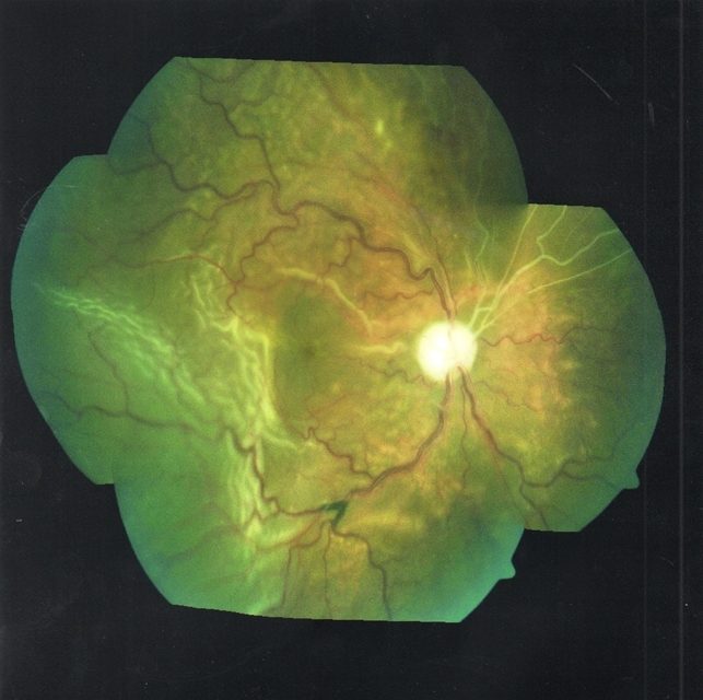
On examination, his best-corrected visual acuity (BCVA) was 20/200 in the right eye and 20/20 in the left eye. Anterior segment examination revealed mild conjunctival injection, while fundus examination demonstrated serous macular detachment, shallow choroidal detachments, and peripapillary serous retinal detachment in the right eye.
The left eye appeared normal. Given the clinical findings, Uveal Effusion Syndrome (UES) was suspected in the right eye.
Uveal Effusion Syndrome (UES) DISEASE entity
Uveal Effusion Syndrome (UES) is a rare syndrome of idiopathic exudative detachments of the choroid, ciliary body, and retina, thought to arise from impaired posterior segment drainage usually associated with scleral thickening.
Idiopathic uveal effusion syndrome usually affects healthy middle-aged men. However, there are no specific risk factors for the disease.
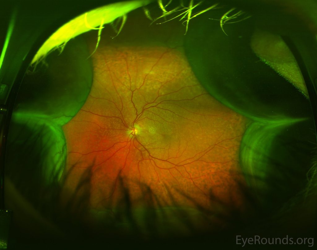
Uveal Effusion Syndrome (UES) was described for the first time in 1963 by RJ Brockhurst. In 1983, JMD Gass hypothesized that the primary underlying cause is a congenital anomaly of the sclera, and in some cases, the vortex veins.
It is sometimes divided into three types:
- Nanophthalmic eyes – type 1. The eyeball is small (average axial length 16 mm) and high hypermetropic (average +16 diopters).
- Non-an ophthalmic eyes with clinically abnormal sclera – type 2. The eyeball size is normal (average axial length 21 mm) with a small refractive error.
- Non-an ophthalmic eyes with clinically normal sclera – type 3.
Fundus Explorer Pro
Photograph the retinal findings described here with the phone already in your pocket — 22 D optics and built-in illumination in one handheld unit.
From Choroida — the team behind this siteMANAGEMENT
Treatment with systemic steroids does not appear to be effective. Surgical decompression of the vortex veins has been described, though the most common treatment is full-thickness sclerotomies to provide choroidal fluid drainage.
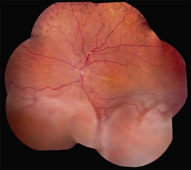
An exit of sub choroidal fluid can be performed by full-thickness sclerectomy, sub scleral sclerectomy, with or without the application of mitomycin C.
Vitrectomy has been described for the management of uveal effusion syndrome. In cases of uveal effusion without an ophthalmic, vitrectomy hastens quick reattachment of the retina and may result in better visual outcomes. In cases of an ophthalmic eye, it would be better to perform a sclerectomy first.
Prognosis:
The largest case series suggests that sclerectomy produces an anatomic improvement in approximately 83% of treated eyes after a single procedure and in about 96% after one or two procedures. Final visual acuity improves by two or more lines in 56% of the eyes, is stable in 35%, and worsens in 9%.
Although extremely rare, UES is a serious condition that is difficult to treat and can lead to severe and permanent visual loss in both eyes.


Document what you see
Two smartphone imaging tools built for everyday clinic use — one for the slit lamp, one for the fundus.
From Choroida — the team behind this siteREFERENCES
- Gass JD. Uveal effusion syndrome. A new hypothesis concerning pathogenesis and technique of surgical treatment.Retina. 1983; 3(3):159-63.
- Uyama M, Takahashi K, Kozaki J, Tagami N, Takada Y, Ohkuma H, Matsunaga H, Kimoto T, Nishimura T. Uveal effusion syndrome: clinical features, surgical treatment, histologic examination of the sclera, and pathophysiology.Ophthalmology. 2000 Mar; 107(3):441-9.
- Ohkita T, Emi K, Toyoda E, Ueno C, Sawada K, Sawada K, Matsumura N, Morita S, Kashimoto D, Oyagi T, Ikeda T. Efficacy of vitreous surgery for uveal effusion syndrome. Nihon Ganka Gakkai Zasshi. 2008 May; 112(5):472-5.
- Elagouz M, Stanescu-Segall D, Jackson TL. Uveal effusion syndrome. Surv Ophthalmol. 2010 Mar-Apr;55(2):134-45.
- Brockhurst RJ. Nanophthalmos with uveal effusion: a new clinical entity. Trans Am Ophthalmol Soc. 1974; 72:371-403.
Test yourself
A few questions straight from this article.
-
Uveal effusion syndrome involves idiopathic exudative detachment of which structures?
UES is a rare idiopathic exudative detachment of the choroid, ciliary body and retina, attributed to impaired posterior segment drainage usually associated with scleral thickening. -
Which patient group does idiopathic uveal effusion syndrome usually affect?
The syndrome typically presents in otherwise healthy middle-aged men, and no specific risk factors for the disease have been identified. -
In nanophthalmic (type 1) uveal effusion syndrome, what are the typical axial length and refraction?
Type 1 nanophthalmic eyes are small, averaging 16 mm axial length, and are highly hypermetropic at around +16 diopters. -
In 1983, Gass proposed which primary underlying cause of uveal effusion syndrome?
Gass hypothesised in 1983 that the primary problem is a congenital scleral anomaly, and in some cases an anomaly of the vortex veins as well. -
Who first described uveal effusion syndrome, and in which year?
RJ Brockhurst gave the first description of uveal effusion syndrome in 1963; Gass later proposed the scleral hypothesis in 1983. -
Which statement describes systemic corticosteroid therapy in uveal effusion syndrome?
Systemic steroids are not effective in UES; treatment is directed instead at creating a route for choroidal fluid to drain through the sclera. -
What is the most common treatment used to drain choroidal fluid in uveal effusion syndrome?
Although vortex vein decompression has been described, the commonest approach is making full-thickness openings in the sclera to allow choroidal fluid to escape. -
After a single scleral drainage procedure for uveal effusion syndrome, what proportion of eyes improve anatomically?
The largest case series reported anatomic improvement in roughly 83 percent of eyes after one procedure, rising to about 96 percent after one or two. -
In treated uveal effusion syndrome, final visual acuity improves by two or more lines in what proportion of eyes?
Final acuity gains of two or more lines occur in 56 percent of eyes, while 35 percent remain stable and 9 percent get worse. -
Which combination of fundus findings fits uveal effusion syndrome?
The affected eye shows serous macular detachment, shallow choroidal detachments and peripapillary serous retinal detachment, while the fellow eye typically looks normal.
