
CASE REPORT
A 32-year-old female presented with progressive vision deterioration for the last 18 months the present status of vision being 20/40(best corrected in both eyes).
General examination and systemic examination were unremarkable. On examination best corrected visual acuity was 20/40 in both eyes, with normal anterior segment examination.
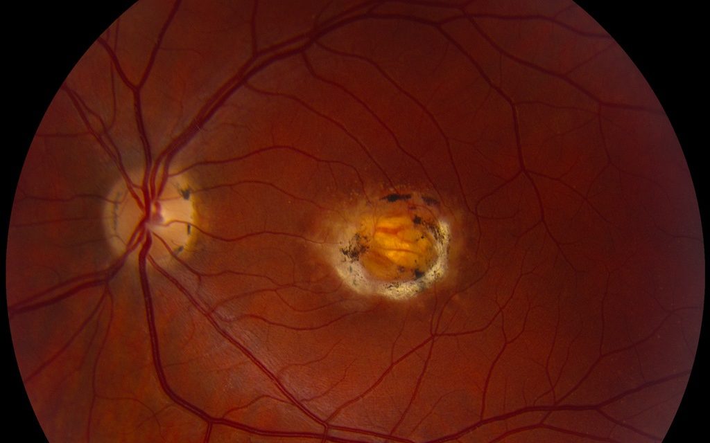
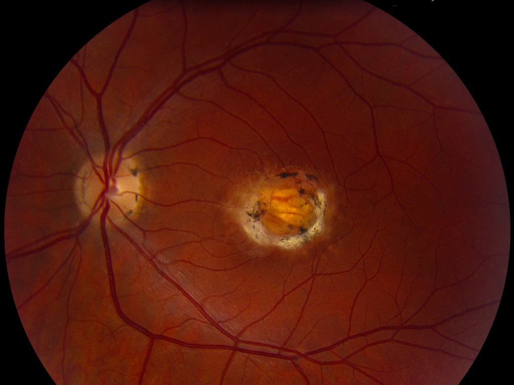
On fundus examination, the posterior pole showed an area of well-delineated chorioretinal degeneration with hyperpigmentation at the border of the lesion. A central crater-like lesion affecting all retinal layers, as well as the deep choroidal tissue, was seen. A provisional diagnosis of North Carolina Macular Dystrophy was made.
North Carolina Macular Dystrophy DISEASE entity
North Carolina macular dystrophy is a congenital, autosomal dominant, non-progressive macular maldevelopment. It is named after a family in North Carolina, who was reported 50 years ago, with congenital, non-progressive macular dystrophy.
These findings were first described by Leffler, Wadsworth, and Sid bury. The macular lesions may evolve until about the age of 12 years, after which they rarely change.
The Macula is an important part of the eye, located in the center of the retina, and provides the sharpest vision. NCMD is a congenitally poor development of the macula.
The symptoms of NCMD vary from patient to patient in accordance with the severity of the disease.
- Some patients are asymptomatic, and some patients complain of mildly to moderate central vision loss. In a few patients, there is a marked impairment of central vision typically due to neovascular membrane formation.
- The color vision remains normal.
- There are no systemic manifestations are associated with North Carolina macular dystrophy.
The disease has been divided into three grades according to severity:
- Grade I: there are fine small/intermediate-sized drusen confining to the central 3° of the central macula.
- Grade II: confluent drusen with or without pigmentary changes.
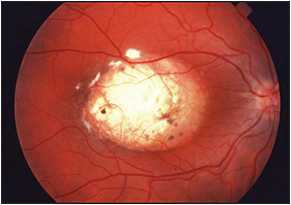
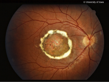
- Grade III: There is well-differentiated chorioretinal atrophy at the macula often surrounding subretinal fibrosis and pigmentation on the edge of the lesion.
Fundus Explorer Pro
Photograph the retinal findings described here with the phone already in your pocket — 22 D optics and built-in illumination in one handheld unit.
From Choroida — the team behind this siteDiagnostic Methods
Clinical examination is performed mainly through the fundus examination on slit lamp biomicroscope and indirect ophthalmoscopy.
Other image modalities like fundus fluorescein angiography, Fundus cameras, Electro retinogram, and Electrooculogram fundus provide further details of the disease.
MANAGEMENT of North Carolina Macular Dystrophy
There should be close monitoring for the development of choroidal neovascular membrane formation. If CNVM develops, intravitreal anti-VEGF injections should be given.
The prognosis is very good. In most cases, the patients have stable vision throughout their life.


Document what you see
Two smartphone imaging tools built for everyday clinic use — one for the slit lamp, one for the fundus.
From Choroida — the team behind this siteREFERENCES
- Rabb MF, Mullen L, Yelchits S, et al. A North Carolina macular dystrophy phenotype in a Belizean family maps to the MCDR1 locus. Am J Ophthalmol. 1998;125:502e508.
- Small K, Small L, Tran E, Rao R, Shaya F. Multimodal Imaging and Functional Testing in a North Carolina Macular Disease Family: Toxoplasmosis, Fovea Plana, and Torpedo Maculopathy Are Phenocopies. Ophthalmology Retina, Volume 3, Issue 7, 607-614.
- Small KW, Vincent A, Knapper CL, Shaya F. Congenital toxoplasmosis as one phenocopy of North Carolina Macular Dystrophy (NCMD/MCDR1). American Journal of Ophthalmology Case Reports, Volume 15, 2019, 100521.
- Small KW, Agemy S, Shaya FS. Terminology of MCDR1: What’s in a name? JAMA Ophthalmol. 2016;134:355e356.
- Benjamin Bakall, MD, PhD, J. Shepard Bryan III, MD, Edwin M. Stone, MD, PhD, Kent W. Small, MD. Choroidal neovascularization in North Carolina macular dystrophy responsive to anti–vascular endothelial growth factor therapy. Retin Cases Brief Rep. 2018 Oct 31. doi: 10.1097/ICB.0000000000000838. PubMed PMID: 30383557.
Test yourself
A few questions straight from this article.
-
What is the inheritance pattern of North Carolina macular dystrophy?
North Carolina macular dystrophy is a congenital, autosomal dominant macular maldevelopment, named after a family from North Carolina. -
What is the underlying nature of North Carolina macular dystrophy?
NCMD is a congenitally poor development of the macula, the central retinal area that provides the sharpest vision. -
How do symptoms vary in North Carolina macular dystrophy?
Symptoms follow disease severity: some patients are asymptomatic, some have mild to moderate central loss, and a few have marked central impairment, typically from a neovascular membrane. -
What happens to colour vision in North Carolina macular dystrophy?
Despite the macular lesion, colour vision remains normal in North Carolina macular dystrophy. -
Which systemic manifestations accompany North Carolina macular dystrophy?
North Carolina macular dystrophy has no associated systemic manifestations; the disease is a macular maldevelopment. -
In grade I North Carolina macular dystrophy, where are the drusen confined?
Grade I shows fine small or intermediate-sized drusen confined to the central 3° of the macula. -
What defines grade III North Carolina macular dystrophy?
Grade III has well-differentiated chorioretinal atrophy at the macula, often with subretinal fibrosis and pigmentation at the lesion edge; drusen define grades I and II. -
How is North Carolina macular dystrophy mainly diagnosed?
Diagnosis is mainly clinical, by fundus examination on the slit lamp biomicroscope and indirect ophthalmoscopy; angiography, fundus photography, ERG and EOG add detail. -
What should patients with North Carolina macular dystrophy be closely monitored for?
Management centres on close monitoring for choroidal neovascular membrane, the usual cause of marked central vision loss in this condition. -
What is the prognosis of North Carolina macular dystrophy?
The prognosis is very good, and in most cases patients keep stable vision throughout their life.
