
CASE REPORT
A 15-year-old child presented with a headache for a few months. His visual acuity came out as 6/6 in both eyes.
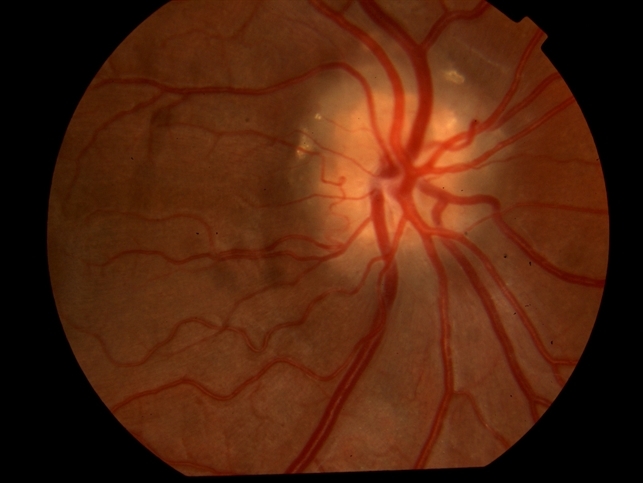
Dilated Fundoscopy revealed elevated discs and blurring of the margins in both eyes. B scan was done to confirm the diagnosis of optic disc drusens.
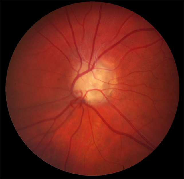
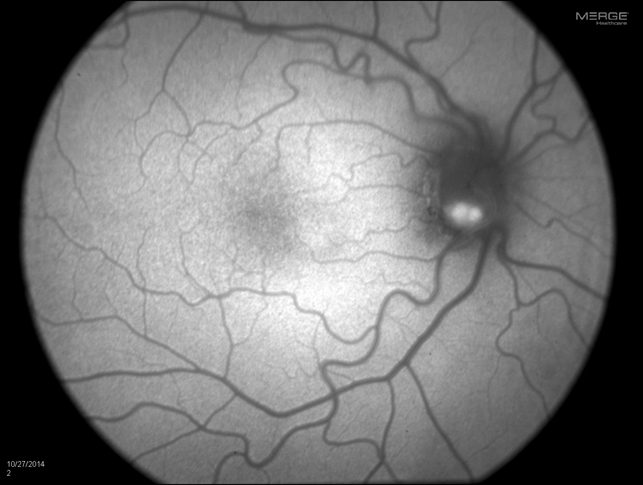
OCT confirmed the thinning of the retinal nerve fiber layer and elevation of the optic disc along with blurry and irregular margins. FAF showed hyper autofluorescence of both discs.
Optic disc drusens entity
Optic disc drusens are progressive, usually bilateral, and asymptomatic types of optic neuropathy in which benign and acellular deposits appear at the optic disc thus making the disc elevated and its margins blurry.
These drusens are mostly mucoproteins or mucopolysaccharides which are Remnants of degenerated retinal ganglion cells. The exact cause of optic disc drusens is not known but is predicted to be due to the abnormal flow of materials in optic nerve cells. There may be a family history of ODD but can also be non-inherited.
These drusens can be classified as buried or superficial. Buried drusen are uncalcified and not seen at the optic nerve head, but the optic disc is elevated with blurry margins due to which they can be misdiagnosed with another term called papilloedema which refers to swelling of the optic disc along with blurring of its margins due to increased intracranial pressure.
Due to their buried nature, it is a challenging task for an ophthalmologist to differentiate between papilloedema and ODD. These are mostly present in children. Superficial drusens are clearly visible at the optic nerve head.
With age optic nerve drusens get calcified and larger, thus compressing normal nerve structures ultimately resulting in visual field defects and vision loss.
Fundus Explorer Pro
Photograph the retinal findings described here with the phone already in your pocket — 22 D optics and built-in illumination in one handheld unit.
From Choroida — the team behind this siteMANAGEMENT of Optic disc drusens
No treatment is available to reverse the visual loss caused by optic disc drusens, although IOP-lowering drugs can be given in patients who have borderline IOP and marked visual field defects.
Complication
They have an increased risk of anterior ischemic optic neuropathy (AION).
Prognosis
ODD usually has a good prognosis for central vision, although visual field defects may progress slowly over time.


Document what you see
Two smartphone imaging tools built for everyday clinic use — one for the slit lamp, one for the fundus.
From Choroida — the team behind this siteREFERENCES
- Thompson AC, Bhatti MT, El-Dairi MA. Bruch’s membrane opening on optical coherence tomography in pediatric papilledema and pseudopapilledema. J AAPOS. 2018;22:38–43.
- Tuğcu B, Özdemir H. Imaging Methods in the Diagnosis of Optic Disc Drusen. Turk J Ophthalmol. 2016;46:232–236.
- Thurtell MJ, Biousse V, Bruce BB, Newman NJ. Optic nerve head drusen in black patients. J Neuroophthalmol. 2012;32:13–16.
- Auw-Haedrich C, Staubach F, Witschel H. Optic disk drusen. Surv Ophthalmol. 2002;47:515–532.
- Lam BL, Morais CG Jr, Pasol J. Drusen of the optic disc. Curr Neurol Neurosci Rep. 2008;8:404–408.
Test yourself
A few questions straight from this article.
-
Which description fits optic disc drusen?
Optic disc drusen are a usually bilateral, asymptomatic optic neuropathy in which benign acellular deposits elevate the disc and blur its margins. -
What are optic disc drusen mainly composed of?
They are mostly mucoproteins or mucopolysaccharides, the remnants of degenerated retinal ganglion cells. -
Which features characterise buried optic disc drusen?
Buried drusen are uncalcified and not seen at the optic nerve head and are mostly present in children, whereas superficial drusen are clearly visible. -
Buried optic disc drusen are most often confused with which condition?
The elevated disc with blurred margins mimics papilloedema, disc swelling caused by raised intracranial pressure, which makes the distinction challenging. -
Which investigation confirmed optic disc drusen in the article's 15-year-old with elevated discs?
After dilated fundoscopy showed elevated discs with blurred margins in both eyes, a B-scan was done to confirm optic disc drusen. -
What did fundus autofluorescence show in the article's case of optic disc drusen?
Fundus autofluorescence showed hyperautofluorescence of both optic discs. -
What did OCT show of the retinal nerve fibre layer in the article's optic disc drusen case?
OCT confirmed retinal nerve fibre layer thinning together with disc elevation and blurred, irregular margins. -
What happens to optic disc drusen with age?
With age the drusen become calcified and larger, compressing normal nerve structures and leading to visual field defects and vision loss. -
When are IOP-lowering drugs given in optic disc drusen?
No treatment reverses the visual loss, but IOP-lowering drugs can be given to patients with borderline IOP and marked visual field defects. -
Which complication carries an increased risk in optic disc drusen?
Patients with optic disc drusen have an increased risk of anterior ischaemic optic neuropathy (AION).
