Tilted disc syndrome (TDS), also called Fuchs coloboma, is a congenital anomaly in which the optic nerve head enters the eye at an oblique angle, usually tilted inferonasally. It affects an estimated 1 to 2% of people. Most patients see well and need no treatment, but the field defects can mimic chiasmal disease.
What is Fuchs coloboma?
Fuchs coloboma is an older name for tilted disc syndrome. The name reflects the inferior thinning, which resembles a coloboma, but the disc is not excavated the way it is in a true optic disc coloboma. The diagnosis often surfaces during a routine dilated exam or while investigating an unexplained field defect.
What does a tilted disc look like on fundus exam?
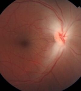
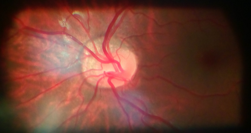
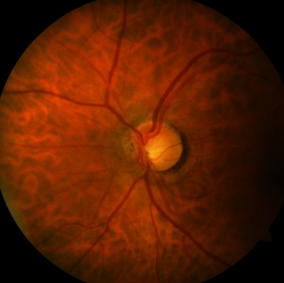
On fundus exam, a tilted disc looks oval, with its long axis rotated away from vertical. The superotemporal edge often looks elevated, while the inferonasal side sits flat or sloped. An inferior or inferonasal crescent borders the disc, and the retina beyond it looks thin and pale.
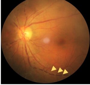
Retinal vessels often leave the disc toward the nose and then curve back, an irregular orientation called situs inversus. Below the disc, the posterior wall of the globe may bulge as an inferior staphyloma, with focal hypopigmentation and thinning of the retinal, choroidal, and scleral layers.
- Oval or obliquely oriented optic disc
- Inferior or inferonasal crescent
- Situs inversus of the retinal vessels
- Inferior or inferonasal ectasia (staphyloma)
- Frequent myopia with astigmatism
Fundus Explorer Pro
Photograph the retinal findings described here with the phone already in your pocket — 22 D optics and built-in illumination in one handheld unit.
From Choroida — the team behind this siteIs a tilted disc the same as a tilted optic disc or oblique insertion?
Yes, in everyday clinical use. Tilted disc, tilted disk, tilted discs, tilted optic disc, and tilted disc syndrome all describe the same finding. Patients who search for “tilted eye syndrome” are asking about it too, although the eye is not tilted. The optic nerve head is.
Oblique insertion describes the anatomy, where the optic nerve and its vessels enter the globe at an angle. Tilted disc describes how that anatomy looks on the fundus. A dragged disc is a different entity, in which traction pulls the nerve head, as in retinopathy of prematurity. Our review of the dragged disc covers the contrast.
What causes tilted disc syndrome?
Tilted disc syndrome is thought to result from oblique insertion of the optic nerve and retinal vessels because the embryonic fissure of the eye closes incompletely. The same process leaves hypoplasia and thinning of the retinal, choroidal, and scleral layers, with ectasia of the inferonasal posterior wall of the globe.
It remains unclear whether the disc anomaly and the inferior staphyloma coexist at birth or whether the staphyloma deepens with time. Most cases are nonhereditary, although autosomal dominant inheritance has been reported. Both eyes are involved in most patients.
How common is tilted disc syndrome?
Tilted disc syndrome occurs in about 1 to 2% of the population and most commonly affects both eyes. Myopia is a frequent companion: about one in five patients with more than 5 diopters of myopia has a tilted disc. Astigmatism is also common and can limit best corrected acuity if left uncorrected.
Because many patients are myopic, a tilted disc is easy to dismiss as a myopic disc. Look at the vessels and the crescent. In TDS the crescent sits inferiorly or inferonasally and the vessels show situs inversus, whereas a typical myopic crescent lies temporally.
Why does tilted disc syndrome cause a visual field defect?
Bitemporal superior visual field defects are present in about one-fifth of patients with tilted discs. The defect corresponds to the thinned, ectatic inferonasal retina, and part of it is a refractive effect from the steep staphyloma. Rucker described these defects in 1944, and later reports called them bitemporal pseudohemianopia.
Unlike chiasmal defects, a tilted disc visual field defect usually does not respect the vertical midline sharply, stays stable, and may shrink when you retest with the best refractive correction. It can also mimic false field deterioration in patients followed for glaucoma or other optic neuropathy, so compare against prior fields before you escalate.
How do you work up a tilted disc visual field defect?
A tilted disc visual field defect calls for a careful but short workup. Start with full refraction and repeat the field with the best correction. Then image the disc and macula. If the defect respects the vertical midline, progresses, or comes with reduced acuity, color loss, or neurologic symptoms, order MRI to exclude a chiasmal lesion.
- Dilated fundus exam documenting the tilt, crescent, and vessel pattern
- Cycloplegic or manifest refraction, including astigmatism
- Repeat perimetry with full correction
- OCT of the macula and optic nerve head
- MRI when the defect has a vertical edge, progresses, or stays unexplained
What else can look like a tilted disc?
A tilted disc can appear elevated and blurred along its superior edge, which sometimes triggers a swelling workup. Check for optic disc drusen with OCT or B-scan ultrasound. Reserve the papilledema pathway for discs with true swelling, such as hyperemia, vessel obscuration, or nerve fiber layer edema.
- Myopic disc with a temporal crescent
- Dragged disc after retinopathy of prematurity
- Optic disc coloboma, with a true excavated defect
- Pseudopapilledema from drusen
Does tilted disc syndrome affect the face?
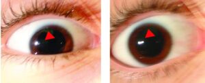
No. Tilted disc syndrome is an ocular finding and causes no facial change. Patients who search for “tilted disc syndrome face” usually want to know whether it signals a wider syndrome. In an otherwise healthy patient with an isolated tilted disc, it does not. In a child with an unusual head or facial shape, consider a broader evaluation.
What are the complications of tilted disc syndrome?
Several macular complications are described in TDS. They include retinal pigment epithelium (RPE) atrophy, choroidal neovascular membrane (CNVM), subretinal fluid (SRF), polypoidal choroidal vasculopathy (PCV), fovea plana, foveoschisis, and lamellar macular hole (LMH). Lacquer cracks have also been reported in a family with dominant inheritance.
Choroidal mechanical and hemodynamic changes at the upper edge of the staphyloma have been proposed to explain these complications. New metamorphopsia, central blur, or a drop in acuity should prompt OCT, and angiography when you suspect CNVM or PCV.


Document what you see
Two smartphone imaging tools built for everyday clinic use — one for the slit lamp, one for the fundus.
From Choroida — the team behind this siteHow do you treat tilted disc syndrome, and what is the prognosis?
The disc itself needs no treatment, and the prognosis is good for most patients. Prescribe full correction with attention to astigmatism, and explain the field findings so patients do not fear a tumor. Record baseline OCT and visual fields so you can compare them at future visits.
Treat complications as you would in any other eye. Active CNVM usually responds to intravitreal anti-VEGF injections, and subretinal fluid without neovascularization needs close follow-up. Because the staphyloma may deepen, schedule periodic dilated exams for patients with high myopia or macular changes, and refer promptly when central symptoms appear.
Test yourself
A few questions straight from this article.
-
What proportion of the population is affected by tilted disc syndrome?
Tilted disc syndrome is a congenital anomaly that occurs in 1 to 2% of the population. -
In which direction is the optic disc characteristically tilted in tilted disc syndrome?
The syndrome is characterised by inferonasal tilting of the optic disc; an inferior or inferonasal crescent is another listed feature. -
How does tilted disc syndrome most commonly present in terms of laterality?
Tilted disc syndrome is a congenital anomaly that most commonly occurs bilaterally. -
What proportion of patients with more than 5 dioptres of myopia have tilted discs?
Tilted disc syndrome is associated with high myopia: one-fifth of patients with more than 5 dioptres of myopia have tilted discs. -
Which visual field defect is present in about one-fifth of patients with tilted discs?
Bitemporal superior visual field defects are found in about one-fifth of patients with tilted discs. -
Which retinal vessel abnormality is a recognised feature of tilted disc syndrome?
Irregular orientation of the retinal vessels (situs inversus) is one of the listed features, along with the inferior crescent and inferior staphyloma. -
What is tilted disc syndrome thought to result from?
It is thought to arise from oblique insertion of the optic nerve and retinal vessels caused by incomplete closure of the embryonic fissure. -
What is known about the inheritance of tilted disc syndrome?
Tilted disc syndrome is mostly understood as a nonhereditary process, although reports of autosomal dominant inheritance exist. -
Which macular complication has been described in tilted disc syndrome?
Described macular complications include CNVM, RPE atrophy, subretinal fluid, polypoidal choroidal vasculopathy, foveoschisis and lamellar macular hole. -
In tilted disc syndrome, where are choroidal changes proposed to drive macular complications?
Mechanical and haemodynamic choroidal changes at the upper edge of the inferior staphyloma have been proposed to explain these macular complications.

Writes and reviews the clinical articles on Educate. Every post is checked against peer-reviewed sources before it goes live.