
CASE REPORT of Ectopia Lentis
A 21-year-old female presented with decreased vision in both eyes (left more than right) noticed for the last year.
Her unaided visual acuity was 20/200 and 20/400 in the right and left eye, respectively, improving to 20/40 with –4.5 DS/–3.0 DC x10° correction in the right eye and 20/80 with –10.0 DS/–6.5 Dc x10° correction in the left eye.
Intraocular pressures were 14 mmHg and 16 mmHg in the right and left eyes, respectively.
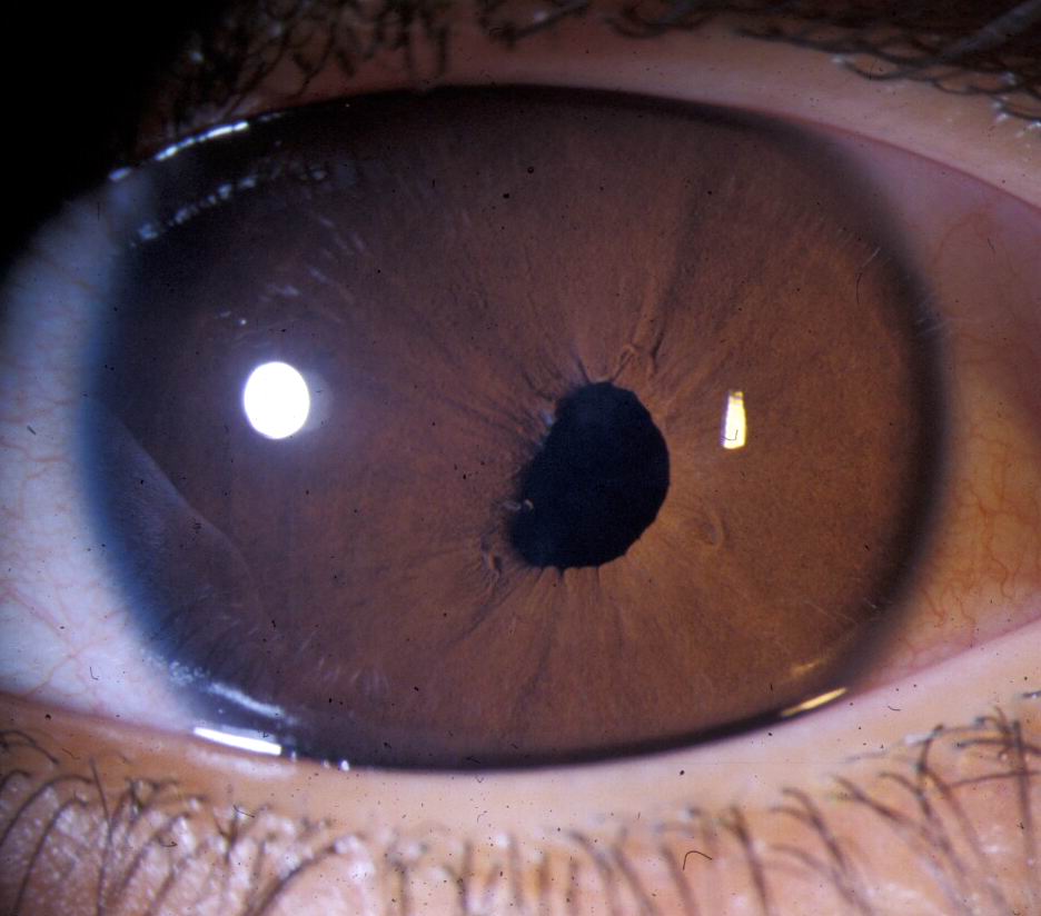
On slit lamp examination, the cornea, iris, anterior chamber, and lens appeared normal in the undilated pupil in both eyes.
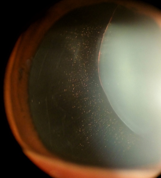
However, on examination under mydriasis, the right eye revealed ectopia lentis.
DISEASE “Ectopia Lentis”
Ectopia lentis is the dislocation or displacement of the natural crystalline lens. The lens is defined as luxated (dislocated) when it lies completely outside of the hyaloid fossa, is free-floating in the vitreous, is in the anterior chamber, or lies directly on the retina.
The lens is considered subluxed when it is partially displaced but remains within the lens space.
Ectopia lentis may occur after trauma or may be associated with ocular or systemic disease. As ectopia lentis may be the first sign of a more serious systemic disease.
it is important to determine the etiology for appropriate patient management.
Additionally,
ectopic lentis can cause major complications within the eye, including significant refractive shifts, pupillary block glaucoma, retinal damage, and blindness.
All-fit Slit-Lamp Adapter
Record and share exactly what you see at the slit lamp. One adapter fits any slit lamp or surgical microscope — and any smartphone.
From Choroida — the team behind this siteEctopia Lentis MANAGEMENT
Medical:
Medical management involves refractive correction as well as co-management of any associated systemic disease with the patient’s internist or pediatrician. As many predisposing diseases are hereditary, genetic counseling may be indicated.
Treatment of ectopia lentis itself depends on where and how far the lens moves, as well as any resulting complications. In all cases, timely optical correction is important for the prevention of amblyopia.
If the dislocated lens migrates into the anterior chamber, treatment is initially pharmacological with mydriasis to permit posterior migration of the lens behind the iris.

Pupillary block glaucoma should be treated with laser peripheral iridotomy, and elevations in intraocular pressure should be treated medically.
Progressive lens dislocation without major complications has been traditionally managed conservatively.
However,
some studies suggest that surgical intervention could lead to improved visual acuity. The decision to operate depends on many factors, including visual acuity, lens location, progressive subluxation of the lens, imminent total dislocation, and others.
Surgical:
Surgical management of ectopia lentis remains a challenge, as inherent defects in the lens capsule make implantation of an intraocular lens difficult.
As a result, many patients are managed with lensectomy/vitrectomy with optical rehabilitation with a contact lens.
Indications for lensectomy include, but are not limited to:
- Lens-induced glaucoma
- Lens in the anterior chamber, especially if the lens is touching the corneal endothelium
- Lens opacity that is mature or hypermature
- Evidence of lens-induced uveitis
- Inadequate visual acuity not correctable by refraction
- Imminent complete luxation of the lens
- Retinal detachment


Document what you see
Two smartphone imaging tools built for everyday clinic use — one for the slit lamp, one for the fundus.
From Choroida — the team behind this siteREFERENCES
- Clarke, Clement C. “Ectopia lentis: a pathologic and clinical study.” Archives of Ophthalmology 21.1 (1939): 124- 153.
- Harley, Robison D., Leonard B. Nelson, and Scott E. Olitsky, eds. Harley’s Pediatric Ophthalmology. Lippincott Williams & Wilkins, 2005. p.225.
- Jarrett, William H. “Dislocation of the lens: a study of 166 hospitalized cases.” Archives of ophthalmology 78.3 (1967): 289-296.
- Chandra, Aman, et al. “A genotype-phenotype comparison of ADAMTSL4 and FBN1 in isolated ectopia lentis.” Investigative ophthalmology & visual science53.8 (2012): 4889-4896.
- Sadiq, Mohammad Ali, and Deborah Vanderveen. “Genetics of ectopia lentis.” Seminars in ophthalmology. Vol. 28. No. 5-6. New York: Informa Healthcare USA, Inc., 2013.
- Nelson, Leonard B., and Irene H. Maumenee. “Ectopia lentis.” Survey of ophthalmology 27.3 (1982): 143-160.
Test yourself
A few questions straight from this article.
-
What does the term ectopia lentis describe?
Ectopia lentis is the dislocation or displacement of the natural crystalline lens from its normal position, whether from trauma or from an underlying ocular or systemic disease. -
In ectopia lentis, when is a lens described as subluxed rather than luxated?
A subluxed lens is only partly displaced and remains within the lens space; a luxated lens has left the hyaloid fossa entirely — floating in vitreous, sitting on the retina, or in the anterior chamber. -
Besides trauma, ectopia lentis is associated with which broad category of cause?
Ectopia lentis may follow trauma or accompany ocular or systemic disease, so the cause must be established before management is planned. -
Why is establishing the etiology of ectopia lentis so important?
A displaced lens can be the presenting sign of a more serious systemic disorder, so identifying the etiology drives appropriate management well beyond the eye. -
Which form of glaucoma is listed as a major complication of ectopia lentis?
Major complications of ectopia lentis include significant refractive shifts, pupillary block glaucoma, retinal damage and ultimately blindness. -
In the reported ectopia lentis case, when did the lens displacement become apparent?
Through the undilated pupil the cornea, iris, anterior chamber and lens all looked normal; the ectopia lentis was revealed only once the pupil was dilated. -
How is a dislocated lens that has migrated into the anterior chamber initially managed?
Treatment begins pharmacologically: the pupil is dilated so the lens can migrate posteriorly behind the iris rather than remain against the corneal endothelium. -
How should pupillary block glaucoma complicating ectopia lentis be treated?
Pupillary block glaucoma is treated with laser peripheral iridotomy, while any accompanying rise in intraocular pressure is managed medically. -
Why is intraocular lens implantation difficult in eyes with ectopia lentis?
The lens capsule itself is defective in these eyes, so an intraocular lens is hard to implant; many patients instead have lensectomy with vitrectomy and are rehabilitated with a contact lens. -
Which finding is a listed indication for lensectomy in ectopia lentis?
Indications for lensectomy include a lens in the anterior chamber, especially where it touches the corneal endothelium, alongside lens-induced glaucoma or uveitis, mature cataract, imminent complete luxation and retinal detachment.
