Uveal melanoma staging follows the AJCC system, which sorts choroidal and ciliary body tumors by size and anatomic extent.
The T category depends on tumor thickness and largest basal diameter, and two modifiers then refine it: ciliary body involvement and extraocular extension. As a result, a few careful measurements carry real prognostic weight.
Why Uveal Melanoma Staging Uses Size
Larger tumors carry a higher risk of metastasis, mainly to the liver, and size is also easy to record at the first visit, before any biopsy. Therefore, the AJCC Ophthalmic Oncology Task Force built the T category around dimensions every center can measure.
Measuring Tumors for Uveal Melanoma Staging
B-scan ultrasonography usually provides thickness, from the inner scleral surface to the apex, and ultrasound and fundus photography together give the largest basal diameter (LBD). Because a dome may hide its true base, scan in several planes, as a tilted probe, for example, can overestimate height.
Fundus Explorer Pro
Photograph the retinal findings described here with the phone already in your pocket — 22 D optics and built-in illumination in one handheld unit.
From Choroida — the team behind this siteThe Size Category Grid
The AJCC table crosses thickness against LBD to give size categories 1 to 4. However, the steps are not symmetric, and a thin but very wide tumor may therefore outrank a thicker, narrower one, and the size category then becomes the T number.
Small Tumors and T1 in Uveal Melanoma Staging
T1 covers small melanomas with modest height and base, for example a tumor up to 3 mm thick with an LBD up to 12 mm, which falls in category 1. Many of these lesions first appear as suspicious nevi (see choroidal nevus vs small melanoma). Small tumors still metastasize in some patients, so small does not mean harmless.
T2 and T3 Tumors
Medium tumors usually fall in T2 or T3, and a tumor between 6 and 9 mm thick with a base up to 12 mm, for instance, is category 2. Thickness above 9 mm generally places a tumor in category 3 or 4. As a result, small errors near a cut point can change the category.
T4 and Very Large Tumors
Any tumor with an LBD greater than 18 mm is category 4, whatever its thickness, and likewise, thickness above 15 mm defines category 4. These eyes often show exudative detachment, secondary glaucoma or a mushroom shape, so enucleation becomes more likely, although selected cases still receive radiotherapy.
Ciliary Body Involvement in Uveal Melanoma Staging
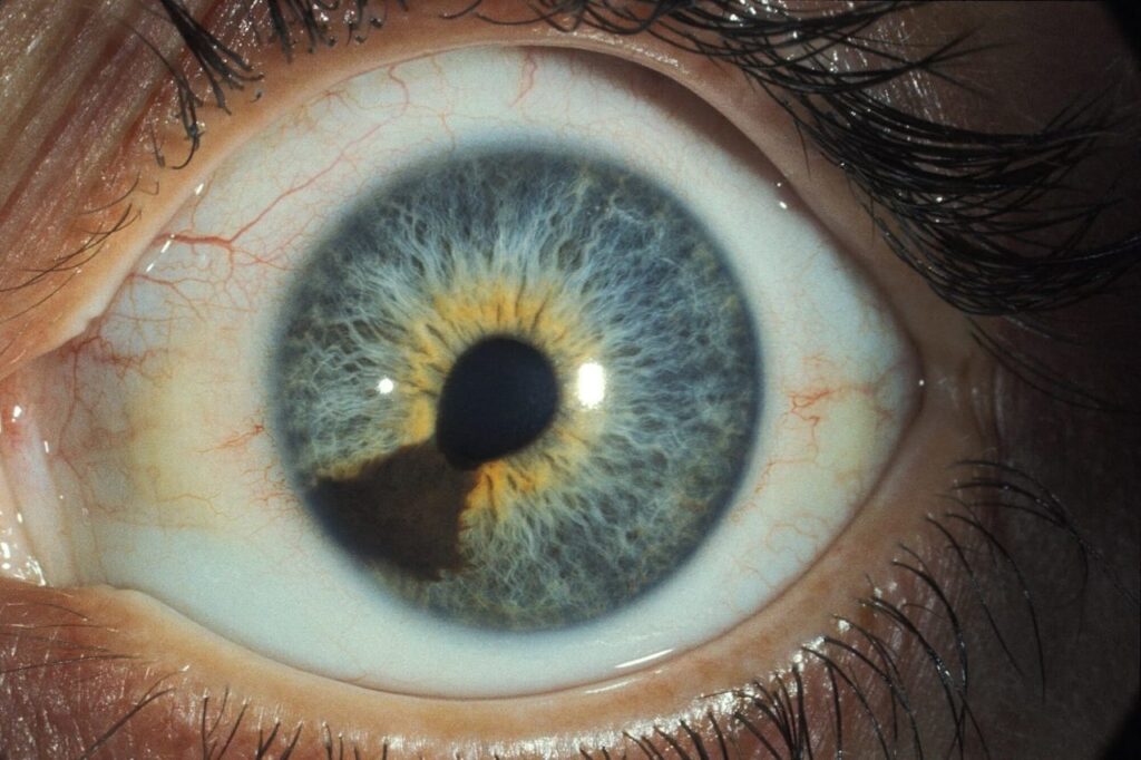
The letter after the T number records anatomic extension, and subcategory “a” means no ciliary body involvement and no extraocular extension. Subcategory “b” adds ciliary body involvement, which worsens prognosis at any size. Because anterior tumors hide behind the iris, use transillumination and ultrasound biomicroscopy (see ciliary body melanoma).
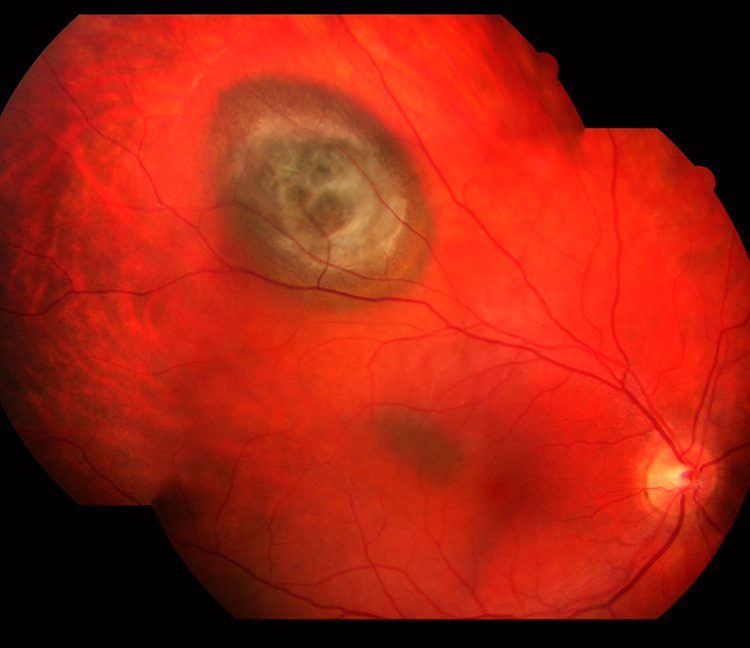
Extraocular Extension Modifiers
Subcategory “c” describes extraocular extension of 5 mm or less without ciliary body involvement, while subcategory “d” combines ciliary body involvement with extension of 5 mm or less. Finally, T4e covers extension greater than 5 mm, regardless of size category, and orbital ultrasound helps detect spread, while pathology confirms it after enucleation.
Uveal Melanoma Staging and Prognosis
Large registry data show metastatic risk rising steadily from T1 to T4, and Shields and colleagues confirmed this pattern in more than 7,000 patients. An international validation study also reported similar trends, and because metastasis can appear many years later, follow-up must stay long.
Anatomic Stage Groups
AJCC then combines T, N and M into stage groups I through IV, and stage I includes only T1a tumors without nodal or distant spread. Higher T subcategories move patients into stages II and III, while any regional node or distant metastasis defines stage IV.
Where Uveal Melanoma Staging Falls Short
Anatomic stage ignores tumor biology, and two tumors of equal size can behave very differently. Monosomy 3, BAP1 loss and a class 2 gene expression profile also add risk information (see uveal melanoma prognostic testing), and histologic cell type carries prognostic value but does not change the T category.
Documenting Uveal Melanoma Staging in the Chart
First, record thickness, LBD, measurement method and margin location, and next, note ciliary body involvement, disc proximity and any extraocular nodule. Then assign the category and letter, and state whether the stage is clinical or pathologic, because clear notes also simplify restaging and trial eligibility.
Counseling Patients About Their Stage
Patients often hear “stage” and assume widespread disease, so explain that most stages describe the tumor inside the eye. Risk rises with size, while genetic results refine the estimate, and a low stage still needs lifelong surveillance.
Pitfalls to Avoid
- Exclude the sclera from thickness measurements
- Never stage from photographs alone when ultrasound is available
- Look for ciliary body involvement in every anterior tumor
- Remember that iris melanoma has its own T system, so the posterior grid does not apply
- Avoid equating a low T category with zero metastatic risk


Document what you see
Two smartphone imaging tools built for everyday clinic use — one for the slit lamp, one for the fundus.
From Choroida — the team behind this siteReferences
- Kivelä T, Simpson ER, Grossniklaus HE, et al. Uveal melanoma. In: Amin MB, ed. AJCC Cancer Staging Manual. 8th ed. Springer; 2017:805-817.
- Shields CL, Kaliki S, Furuta M, et al. American Joint Committee on Cancer classification of posterior uveal melanoma (tumor size category) predicts prognosis in 7731 patients. Ophthalmology. 2013;120:2066-2071.
- AJCC Ophthalmic Oncology Task Force. International validation of the American Joint Committee on Cancer’s 7th edition classification of uveal melanoma. JAMA Ophthalmol. 2015;133:376-383.
- Kujala E, Mäkitie T, Kivelä T. Very long-term prognosis of patients with malignant uveal melanoma. Invest Ophthalmol Vis Sci. 2003;44:4651-4659.

Writes and reviews the clinical articles on Educate. Every post is checked against peer-reviewed sources before it goes live.