
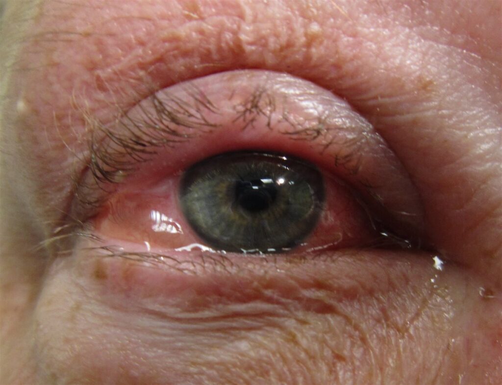
One symptom, more than any other, separates allergy from infection when a patient walks in with red, watery eyes.
Allergic conjunctivitis is itching — genuine, hard-to-ignore itching — and its presence or absence changes the entire diagnostic conversation.
It is among the most common ocular surface conditions seen in both primary care and ophthalmology.
Most cases are mild and self-limited, but a smaller subset — vernal and atopic keratoconjunctivitis — can threaten the cornea and vision itself.
Patients are frequently treated with antibiotic drops for what is actually an allergic, not infectious, process.
Correctly classifying the type of allergic conjunctivitis present is what determines whether simple avoidance and antihistamines are enough, or whether more aggressive anti-inflammatory treatment is needed.
What Is Allergic Conjunctivitis?
Allergic conjunctivitis is an IgE-mediated hypersensitivity reaction of the conjunctiva to environmental allergens.
It spans a spectrum of severity, classified as:
- Seasonal allergic conjunctivitis (SAC) — triggered by airborne pollens, flaring with the seasons
- Perennial allergic conjunctivitis (PAC) — triggered by year-round allergens such as dust mites and animal dander
- Vernal keratoconjunctivitis (VKC) — a more severe, chronic form, most common in boys and young men with a personal or family history of atopy
- Atopic keratoconjunctivitis (AKC) — a severe, chronic form associated with atopic dermatitis, typically in adults
- Giant papillary conjunctivitis (GPC) — a mechanically driven variant related to contact lens wear or an ocular prosthesis
Seasonal and perennial disease account for the great majority of cases and are generally mild; VKC and AKC are far less common but carry real risk to the cornea.
Epidemiology
Allergic conjunctivitis is extremely common and closely tracks the broader burden of allergic disease.
- Seasonal and perennial allergic conjunctivitis together account for the large majority of allergic conjunctivitis cases
- Vernal keratoconjunctivitis is most common in boys and young men aged roughly 5-20 years, often with coexisting eczema, asthma, or seasonal allergies
- Atopic keratoconjunctivitis typically presents later, in adults with a background of atopic dermatitis
- Prevalence is rising alongside the broader increase in allergic and atopic disease worldwide
Because most patients self-treat mild seasonal symptoms without ever seeking care, true prevalence is almost certainly higher than clinic-based figures suggest.
All-fit Slit-Lamp Adapter
Record and share exactly what you see at the slit lamp. One adapter fits any slit lamp or surgical microscope — and any smartphone.
From Choroida — the team behind this sitePathophysiology
The underlying mechanism is a classic type I hypersensitivity reaction, more pronounced in the severe chronic forms.
- Allergen exposure cross-links IgE bound to conjunctival mast cells
- Mast cell degranulation releases histamine and other inflammatory mediators
- Histamine release produces the itching, redness, and watery discharge typical of acute allergic conjunctivitis
- In VKC and AKC, chronic T-cell-mediated inflammation adds a second, more destructive layer on top of the acute mast cell response
- Chronic inflammation in these severe forms can produce giant papillae and, in advanced disease, corneal epithelial damage
This is why simple antihistamines control most seasonal and perennial disease, but VKC and AKC often need broader anti-inflammatory treatment aimed at the chronic T-cell component.
Risk Factors
Patient-Related Risk Factors
- A personal or family history of atopy — asthma, eczema, or allergic rhinitis
- Young age and male sex, particularly for vernal keratoconjunctivitis
- Living in warm, dry climates, associated with a higher prevalence of vernal disease
Exposure-Related Risk Factors
- Seasonal pollen exposure for SAC
- Dust mites, animal dander, and indoor allergens for PAC
- Contact lens wear, a specific risk factor for giant papillary conjunctivitis
Identifying the specific trigger, when possible, is what makes allergen avoidance a genuinely useful part of management rather than a vague recommendation.
Clinical Presentation
Symptoms
- Itching — the hallmark symptom, and the single best clue that an allergic rather than infectious process is at work
- Watery discharge, in contrast to the purulent discharge of bacterial conjunctivitis
- Bilateral involvement in the great majority of cases
- Associated allergic symptoms such as sneezing or nasal congestion in many patients
Examination Findings
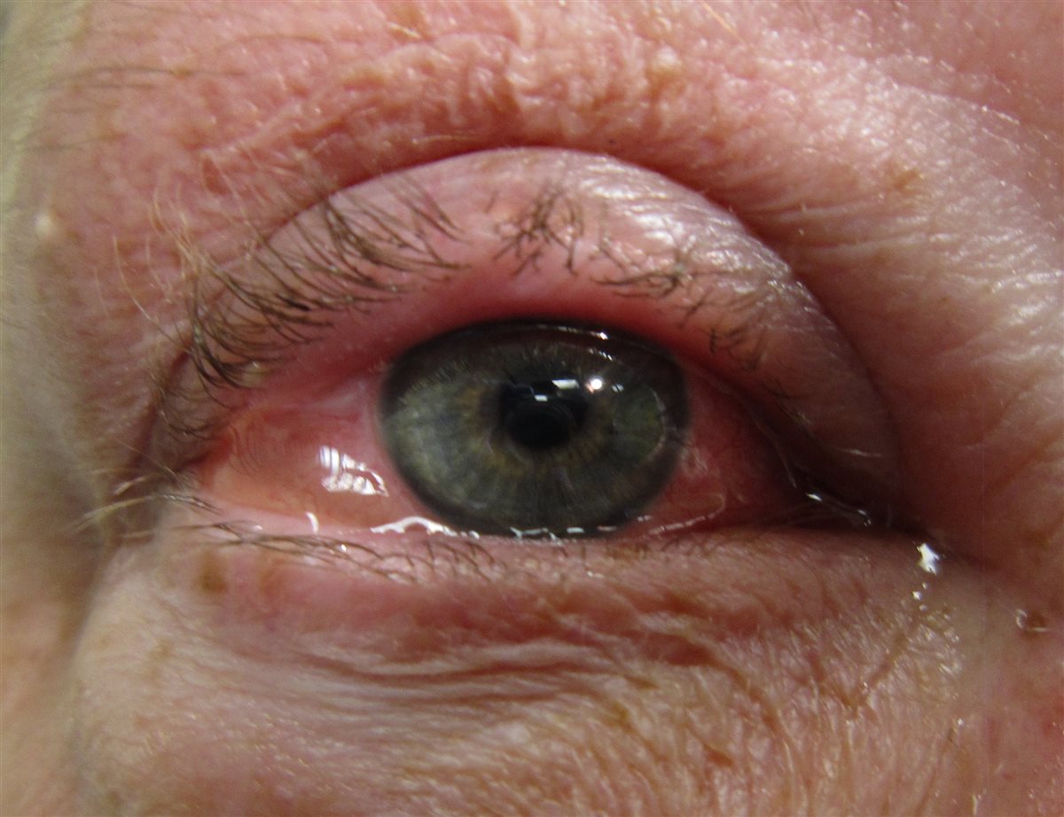
- Diffuse conjunctival injection with a clear, watery discharge
- Conjunctival chemosis (edema) in more acute or severe presentations
- Mild papillary reaction on the tarsal conjunctiva in simple SAC/PAC
- Giant papillae (“cobblestoning”) of the upper tarsal conjunctiva in vernal keratoconjunctivitis
- Corneal involvement — punctate epithelial erosions or, in severe VKC, a shield ulcer — signals disease severe enough to threaten vision
Bilateral itching with a watery, non-purulent discharge is usually enough to make the diagnosis without further testing in straightforward cases.
Diagnostic Evaluation
Clinical Examination
- Diagnosis is primarily clinical, based on the combination of itching, bilateral injection, and watery discharge
- Eyelid eversion to assess for papillae, particularly when VKC, AKC, or GPC is suspected
Assessing Severity
- Fluorescein staining of the cornea to identify punctate erosions or a shield ulcer in suspected vernal or atopic disease
- Assessment of associated eyelid skin changes in suspected atopic keratoconjunctivitis
Allergy Workup
- Formal allergy testing is not routinely required but can help identify specific triggers in patients with persistent or poorly controlled symptoms
- A directed history of seasonal pattern, pets, and occupational exposures is often more immediately useful than formal testing
Corneal involvement is the key finding that separates a condition needing reassurance and drops from one needing close, active management.
Differential Diagnosis
Conditions that can be confused with allergic conjunctivitis include:
- Viral conjunctivitis — watery discharge can overlap, but itching is far less prominent and preauricular adenopathy is common
- Bacterial conjunctivitis — purulent rather than watery discharge, and typically unilateral at onset
- Dry eye disease — burning and foreign-body sensation predominate over true itching
- Blepharitis — lid margin crusting and irritation without the acute bilateral itching of allergic conjunctivitis
- Contact dermatitis of the eyelids — periocular skin changes from a topical exposure, sometimes coexisting with allergic conjunctivitis
Itching remains the single best discriminator — its presence points strongly toward allergy, and its absence should prompt reconsideration of an infectious cause.
Management
Seasonal and Perennial Disease
- Allergen avoidance where practical — reducing exposure to the identified or suspected trigger
- Cold compresses and preservative-free artificial tears for symptomatic relief
- Topical dual-acting antihistamine/mast cell stabilizer drops as first-line pharmacologic therapy
- Oral antihistamines as an adjunct, particularly with coexisting allergic rhinitis
Vernal and Atopic Keratoconjunctivitis
- Topical antihistamine/mast cell stabilizer therapy remains foundational, as in milder disease
- Topical corticosteroids for acute exacerbations, used for the shortest effective course given the risks of prolonged steroid use
- Topical calcineurin inhibitors (e.g., cyclosporine) as steroid-sparing agents for chronic control
- Close monitoring for corneal involvement, since a shield ulcer or significant epithelial disease requires more urgent, targeted treatment
Giant Papillary Conjunctivitis
- Discontinuing or modifying the mechanical trigger — contact lens wear, lens material, or wear schedule
- Anti-inflammatory drops as needed for symptomatic control
Escalating treatment should be reserved for the minority of patients with VKC, AKC, or corneal involvement — most allergic conjunctivitis responds well to simple, low-risk measures.
Prognosis
Prognosis is excellent for the great majority of patients with seasonal or perennial disease.
- SAC and PAC typically respond well to avoidance measures and topical antihistamine/mast cell stabilizer therapy, with no lasting ocular damage
- VKC often improves after puberty, though it can cause corneal scarring and visual impairment if poorly controlled during active disease
- AKC follows a more chronic course and carries a higher risk of long-term corneal complications, including scarring and secondary infection
Long-term visual outcome depends almost entirely on whether corneal involvement is identified and treated early in the more severe subtypes.
Would you like to document anterior segment findings with your smartphone?


Document what you see
Two smartphone imaging tools built for everyday clinic use — one for the slit lamp, one for the fundus.
From Choroida — the team behind this siteReferences
- American Academy of Ophthalmology. Conjunctivitis Preferred Practice Pattern. 2024.
- Allergic Conjunctivitis. Merck Manual, Professional Edition.
- Bielory L, Delgado L, Katelaris CH, et al. ICON: Diagnosis and management of allergic conjunctivitis. Annals of Allergy, Asthma & Immunology. 2020.
- Allergic Conjunctivitis. EyeWiki, American Academy of Ophthalmology.
- Leonardi A, Doan S, Fauquert JL, et al. Diagnostic tools in ocular allergy. Allergy. 2017.
Test yourself
A few questions straight from this article.
-
Which symptom is the hallmark that separates allergic conjunctivitis from an infectious cause?
Itching is the single best discriminator. Its presence points strongly toward allergy, and its absence should prompt reconsideration of an infectious cause. -
What is the underlying immune mechanism of allergic conjunctivitis?
Allergen exposure cross-links IgE bound to conjunctival mast cells, and degranulation releases histamine and other mediators that produce itching, redness and watery discharge. -
Which subtype of allergic conjunctivitis is mechanically driven by contact lens wear?
Giant papillary conjunctivitis is a mechanically driven variant related to contact lens wear or an ocular prosthesis, managed by changing or stopping the trigger. -
Which patient group is most typically affected by vernal keratoconjunctivitis?
Vernal keratoconjunctivitis is most common in boys and young men roughly 5-20 years old, often with coexisting eczema, asthma or seasonal allergies. -
Which tarsal conjunctival finding is characteristic of vernal keratoconjunctivitis?
Chronic inflammation in vernal disease produces giant papillae — cobblestoning — on the upper tarsal conjunctiva, which is why eyelid eversion belongs in the exam. -
Which corneal finding signals vernal keratoconjunctivitis severe enough to threaten vision?
Corneal involvement — punctate epithelial erosions or, in severe vernal disease, a shield ulcer — marks disease severe enough to threaten vision and needs urgent targeted treatment. -
What is first-line pharmacologic therapy for seasonal and perennial allergic conjunctivitis?
Dual-acting antihistamine/mast cell stabilizer drops are first-line, alongside allergen avoidance, cold compresses and preservative-free artificial tears. -
Which agents are used as steroid-sparing therapy for chronic vernal or atopic keratoconjunctivitis?
Calcineurin inhibitors give chronic control while limiting the risks of prolonged steroid use, which is reserved for the shortest effective course in acute exacerbations. -
Which feature points to viral rather than allergic conjunctivitis?
Watery discharge overlaps between the two, but in viral conjunctivitis itching is far less prominent and preauricular adenopathy is common. -
What is the typical long-term course of vernal keratoconjunctivitis?
Vernal disease often improves after puberty, though poorly controlled active disease can still cause corneal scarring and visual impairment along the way.

Writes and reviews the clinical articles on Educate. Every post is checked against peer-reviewed sources before it goes live.


