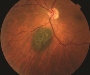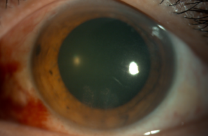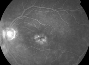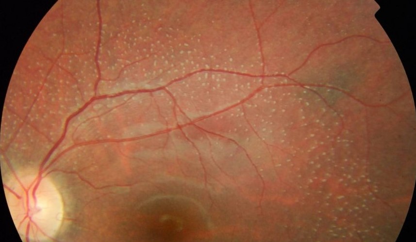
Retinitis punctata albescens is a progressive retinal dystrophy that combines small white dots with signs of retinitis pigmentosa.
Patients have night blindness from early life, and many then develop macular disease in adulthood.
However, it must be separated from fundus albipunctatus, which is stationary and has a different genetic basis.
What Retinitis Punctata Albescens Is
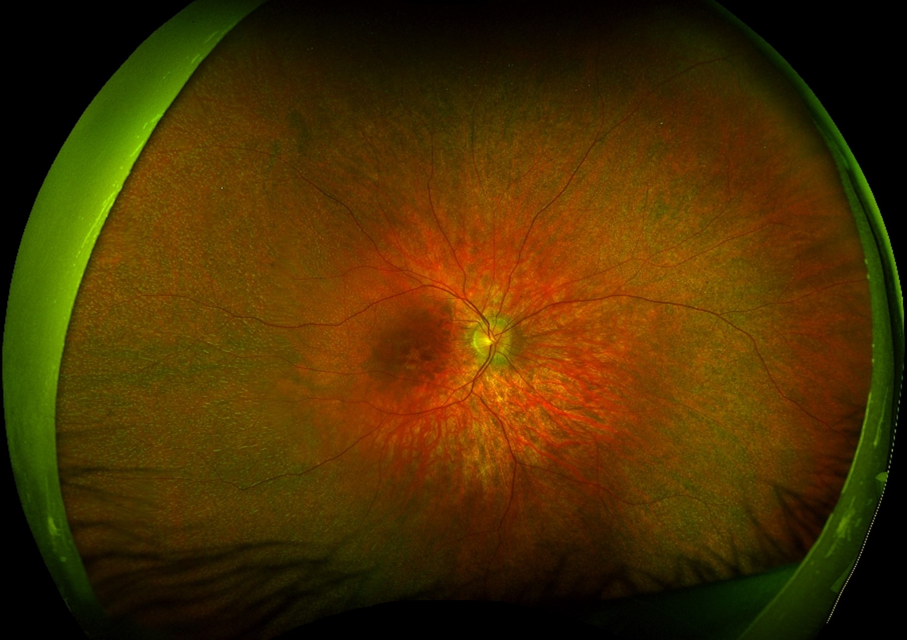
First, the disease shows hundreds of small white or yellow dots at the level of the RPE in the mid-periphery.
Then, over time, the retina develops vessel attenuation, optic disc pallor, and pigment changes.
The condition is a variant of retinitis pigmentosa with a distinct fundus appearance (see retinitis pigmentosa).
Clinical Features of Retinitis Punctata Albescens
- Night blindness from childhood
- Progressive field constriction and loss of central vision
- White dots that may fade as disease advances
- Retinal vessel narrowing and mild pigment changes
Also, some patients have reduced central vision from macular involvement before middle age.
Fundus Explorer Pro
Photograph the retinal findings described here with the phone already in your pocket — 22 D optics and built-in illumination in one handheld unit.
From Choroida — the team behind this siteGenetics of Retinitis Punctata Albescens
Mutations in RLBP1, which encodes cellular retinaldehyde-binding protein, cause Bothnia dystrophy and some forms of retinitis punctata albescens.
Maw and colleagues reported the first mutation in a family with recessive disease.
Burstedt and colleagues described the Bothnia founder variant in northern Sweden.
Also, other genes include RHO and PRPH2 in dominant forms.
Retinitis Punctata Albescens Versus Fundus Albipunctatus
In contrast, fundus albipunctatus is stationary and has normal fields, with RDH5 mutations.
However, retinitis punctata albescens progresses, has constricted fields, and shows vessel attenuation.
Dark adaptation extended to many hours may normalize ERG in albipunctatus but not in the progressive disorder (see fundus albipunctatus).
Investigations for Retinitis Punctata Albescens
ERG shows reduced rod and cone responses, often with worse rod function.
OCT shows outer retinal thinning and, in some patients, macular cystic changes.
Genetic testing confirms the diagnosis and informs counseling (see electroretinography).
Management of Retinitis Punctata Albescens
Because there is no specific therapy, care follows the principles of retinitis pigmentosa.
Then treat macular edema, advise sun protection, and offer low vision support.
Provide genetic counseling to affected families.
An Example Case
A 17-year-old boy reports poor vision at night since childhood, and the fundus shows many fine white dots in the mid-periphery and narrow vessels.
His fields are constricted to 30 degrees, and ERG shows reduced rod and cone responses.
Genetic testing finds biallelic RLBP1 mutations, and the diagnosis is retinitis punctata albescens.
The family receives counseling, and he begins visual rehabilitation.
Questions Patients Ask
Patients ask whether the white dots are dangerous, and they are a marker, and not the cause, of vision loss.
They also ask whether the disease will progress, and the progressive form usually worsens over decades.
Because night blindness affects safety, offer practical advice on lighting and travel.
Pitfalls to Avoid
- Confusing the condition with the stationary fundus albipunctatus
- Skipping ERG after a long dark adaptation
- Missing macular edema that responds to treatment
- Forgetting genetic counseling for siblings
Looking Ahead
Gene panels now make the diagnosis easier, and trials for retinitis pigmentosa may widen to other genes.
Also, better imaging may show the dots on autofluorescence.
Therefore, patients may soon access more options.
Practical Points
Order full-field ERG with long dark adaptation.
Then add OCT, fields, and genetic testing.
Also coordinate with a low vision service.
Autofluorescence Findings
Dots may appear bright or dark on autofluorescence, depending on the stage.
The central macula may show a ring of increased signal in some patients.
Therefore, use autofluorescence to track change.
Family Counselling
Most forms follow recessive inheritance, so siblings have a 25 percent chance of disease.
Dominant forms affect a parent in each generation.
Because inheritance matters, document the pedigree.
Dark Adaptation Testing
Long dark adaptation of two hours or more helps to separate the stationary and progressive forms.
If rod function normalizes, fundus albipunctatus is likely.
However, if it stays low, think of a progressive dystrophy.
Therefore, test with patience and good technique.
Macular Edema and Treatment
Cystoid macular edema occurs in some patients and may respond to carbonic anhydrase inhibitors.
Also consider topical or periocular therapy in selected eyes.
Because edema reduces vision beyond the dystrophy itself, check OCT at each visit.
Summary
White dots with progressive night blindness define the disorder.
Therefore, perform ERG with long dark adaptation and genetic testing.
Because treatment is limited, focus on counseling and rehabilitation.
For the latest studies, see the PubMed results for retinitis punctata albescens.


Document what you see
Two smartphone imaging tools built for everyday clinic use — one for the slit lamp, one for the fundus.
From Choroida — the team behind this siteReferences
- Maw MA, Kennedy B, Knight A, et al. Mutation of the gene encoding cellular retinaldehyde-binding protein in autosomal recessive retinitis pigmentosa. Nat Genet. 1997;17:198-200.
- Burstedt MS, Sandgren O, Holmgren G, Forsman-Semb K. Bothnia dystrophy caused by mutations in the cellular retinaldehyde-binding protein gene (RLBP1) on chromosome 15q26. Invest Ophthalmol Vis Sci. 1999;40:995-1000.
- Hartong DT, Berson EL, Dryja TP. Retinitis pigmentosa. Lancet. 2006;368:1795-1809.
- American Academy of Ophthalmology. Basic and Clinical Science Course, Section 12: Retina and Vitreous. AAO; current edition.

Writes and reviews the clinical articles on Educate. Every post is checked against peer-reviewed sources before it goes live.