Immunosuppression after keratoplasty protects the graft from the host immune system.
The cornea is an immune-privileged site, and most low-risk grafts survive with topical steroids alone.
In high-risk eyes, the drops are not enough, and systemic drugs may be needed.
Why Immunosuppression After Keratoplasty Is Needed
The normal cornea lacks blood vessels and lymphatics, and the anterior chamber has features that reduce immune responses, which together give the graft a good start (see the references).
When these features are lost, as with vascularization, inflammation, or a repeat graft, the immune system can recognize the donor tissue and attack it (see corneal graft rejection).
Endothelial rejection is the most dangerous form, because the endothelium cannot regenerate.
Immunosuppression After Keratoplasty: Who Is at High Risk?
- Corneas with two or more quadrants of deep vascularization
- Repeat grafts after a previous rejection
- Herpetic keratitis or active inflammation at the time of surgery
- Young recipients, particularly children
- Large and eccentric grafts that lie close to the limbus
- Ocular surface disease, such as chemical injury or limbal stem cell deficiency (see limbal stem cell deficiency)
Low-risk grafts, such as those for keratoconus in a healthy eye, have a rejection rate of only a few percent.
All-fit Slit-Lamp Adapter
Record and share exactly what you see at the slit lamp. One adapter fits any slit lamp or surgical microscope — and any smartphone.
From Choroida — the team behind this siteTopical Corticosteroids
Topical steroids are the foundation of treatment.
- Prednisolone acetate 1 percent is started frequently after surgery, often every two hours while awake, and tapered over weeks
- In penetrating grafts, a low dose is continued for 6 to 12 months, and sometimes for years in high-risk cases
- After endothelial keratoplasty, the rejection rate is lower, particularly after Descemet membrane endothelial keratoplasty, and the steroid course can be shorter (see endothelial keratoplasty)
- Monitor the intraocular pressure, since steroid response is common (see steroid-induced glaucoma)
Cataract is a long-term risk of steroid use.
Immunosuppression After Keratoplasty: Systemic Immunosuppression
Clinicians consider systemic drugs in high-risk grafts, and in eyes that have rejected despite topical therapy.
- Tacrolimus: a calcineurin inhibitor with a good record in high-risk grafts
- Mycophenolate mofetil: an antimetabolite that has reduced rejection in randomized and observational studies (see the references)
- Cyclosporine: used orally, and topically in some regimens
- Systemic steroids: used for short courses in an acute rejection
These drugs need blood tests, a physician familiar with transplant medicine, and a discussion of the risks, which include infection, kidney problems, and blood count changes.
HLA Matching
The Collaborative Corneal Transplantation Studies tested tissue matching in high-risk grafts and found that HLA matching did not improve survival, while a low ABO mismatch and good crossmatching offered little benefit in routine practice (see the references).
For this reason, matching is not widely used.
Immunosuppression After Keratoplasty: Monitoring for Rejection
Tell patients the warning signs, which are redness, light sensitivity, reduced vision, and pain.
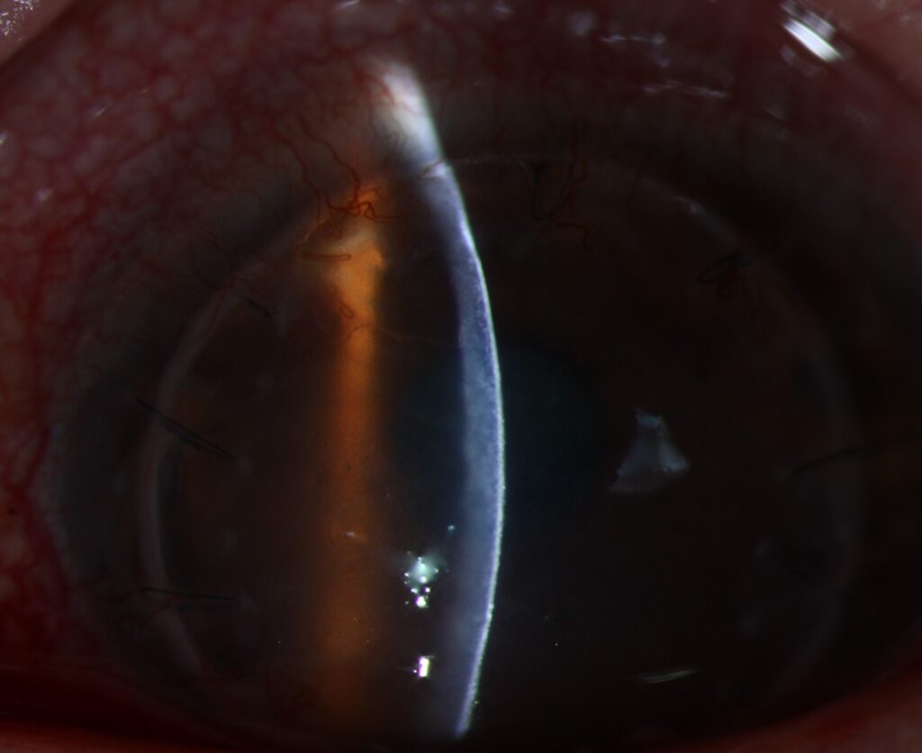
Signs at the slit lamp include keratic precipitates on the graft, a rejection line, stromal edema, and anterior chamber reaction.
Treatment of an episode is hourly steroids, often with a short oral or intravenous course in severe cases, and prompt review.
Practical Pointers
- Educate the patient before the graft, and give a written plan
- Do not stop steroid drops without advice
- Review in the clinic at regular intervals, with pressure checks
- For patients on systemic drugs, coordinate care with a transplant or rheumatology physician
An Example of a Stepwise Plan
Consider a regraft in a vascularized cornea after a first graft failed from rejection.
The surgeon starts frequent topical steroids at surgery and continues a long taper, adds topical cyclosporine or tacrolimus, and arranges blood tests and a discussion with a transplant physician about oral tacrolimus or mycophenolate for the first year.
The patient is seen at close intervals, with pressure checks and a written plan for symptoms.
Special Situations
- Herpetic keratitis: use oral antiviral prophylaxis alongside steroids, since the virus may reactivate after surgery (see herpes simplex keratitis)
- Pediatric grafts: rejection is frequent and quick to progress, so frequent review under anesthesia may be needed
- Endothelial keratoplasty: a lower steroid dose may be used, with attention to pressure
- Limbal stem cell transplantation: systemic immunosuppression is usually needed for allografts
Safety of Systemic Drugs
Patients on tacrolimus need monitoring of kidney function, blood pressure, and drug levels, and those on mycophenolate need blood counts.
Both drugs raise the risk of infection and, with long-term use, of some tumors, so the decision is individual.
Clinicians usually consider stopping after one to two years if the graft is stable.
For the latest studies, see the PubMed results for immunosuppression after keratoplasty.


Document what you see
Two smartphone imaging tools built for everyday clinic use — one for the slit lamp, one for the fundus.
From Choroida — the team behind this siteReferences
- Niederkorn JY. Corneal transplantation and immune privilege. Int Rev Immunol. 2013;32:57-67.
- Collaborative Corneal Transplantation Studies Research Group. The Collaborative Corneal Transplantation Studies (CCTS): effectiveness of histocompatibility matching in high-risk corneal transplantation. Arch Ophthalmol. 1992;110:1392-1403.
- Reinhard T, Mayweg S, Sokolovska Y, et al. Systemic mycophenolate mofetil avoids immune reactions in penetrating high-risk keratoplasty: preliminary results of an ongoing prospectively randomized multicentre study. Transpl Int. 2005;18:703-708.
- American Academy of Ophthalmology. Basic and Clinical Science Course, Section 8: External Disease and Cornea. AAO; current edition.

Writes and reviews the clinical articles on Educate. Every post is checked against peer-reviewed sources before it goes live.