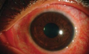
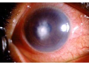
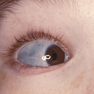
Thalassemia retinopathy is the group of retinal changes found in patients with beta thalassemia, especially those who need regular blood transfusions.
The fundus changes are often subtle, and most patients keep good vision, but they reflect the effects of chronic anemia, iron overload, and treatment.
An ophthalmologist who knows the pattern can also separate it from other causes of retinal pigment changes.
What Causes Thalassemia Retinopathy?
Beta thalassemia is an inherited reduction in the production of beta globin chains, which leads to ineffective red blood cell production and chronic hemolytic anemia.
Also, patients with thalassemia major depend on transfusions, and iron accumulates in tissues, including the retinal pigment epithelium and the choroid.
Chronic anemia and hypoxia may also contribute to retinal changes.
Some patients also show an elastic tissue defect similar to pseudoxanthoma elasticum, which affects Bruch membrane (see pseudoxanthoma elasticum).
In addition, iron chelation therapy has its own effects.
The drug deferoxamine has been linked to retinal toxicity in some patients.
Clinical Findings
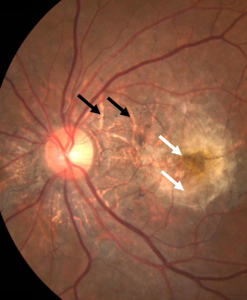
- Retinal pigment epithelial changes: mottling, stippling, and patches of atrophy, especially in the posterior pole and midperiphery
- Angioid streaks, which are also cracks in Bruch membrane that radiate from the optic disc (see angioid streaks)
- Drusen-like deposits in the macula in some patients
- Venous tortuosity and dilation, and occasionally retinal hemorrhages
- Peripapillary and macular atrophy in advanced cases
- Choroidal neovascularization, in rare cases with angioid streaks
- Optic disc changes, in a few patients
The changes are more frequent in older patients and in those with a longer disease duration and greater iron load.
Fundus Explorer Pro
Photograph the retinal findings described here with the phone already in your pocket — 22 D optics and built-in illumination in one handheld unit.
From Choroida — the team behind this siteFrequency and Visual Function
Studies report retinal abnormalities in a substantial proportion of patients with thalassemia major, with angioid streaks reported in a minority and pigment changes in many.
Visual acuity is normal in most patients, and the risk of vision loss from the retinal disease is low.
Complications such as macular hemorrhage and choroidal neovascularization can reduce vision, and they need prompt attention.
Differential Diagnosis of Thalassemia Retinopathy
- Sickle cell retinopathy, with peripheral changes and neovascularization (see sickle cell retinopathy)
- Pseudoxanthoma elasticum and other causes of angioid streaks, including Paget disease and Ehlers-Danlos syndrome (see Ehlers-Danlos syndrome)
- Hemosiderosis and siderosis from other causes (see siderosis bulbi)
- Drug toxicity, including deferoxamine retinopathy (see deferoxamine retinopathy)
- Hypertensive and other vascular retinopathies
- Age-related macular degeneration, in older patients
A history of thalassemia and the pattern of the changes usually identify the cause.
Evaluation for Thalassemia Retinopathy
- Fundus examination with dilation and photographs
- OCT of the macula, which can show pigment epithelial changes, drusen-like deposits, and fluid
- Fundus autofluorescence for pigment epithelial damage
- Fluorescein angiography and OCT angiography if neovascularization is suspected (see OCT angiography)
- Review of the iron burden, chelation regimen, and any visual or hearing symptoms with the hematologist
Electroretinography may be abnormal in some patients with toxicity from chelators.
Screening
Patients with thalassemia should have a baseline eye examination and regular follow-up, often yearly, with more frequent review if they are on deferoxamine or have visual symptoms.
A patient who notices night blindness, reduced vision, or color change needs prompt assessment, since toxicity from chelators can be reversible if it is detected early.
Management for Thalassemia Retinopathy
There is no specific treatment for the retinal pigment changes themselves.
Good control of anemia and iron overload is important for the overall health of the patient.
The hematologist chooses the chelator, with ophthalmic monitoring.
Clinicians treat neovascular complications with anti-VEGF injections (see anti-VEGF therapy).
Counsel patients that most retinal changes do not affect vision, and that regular follow-up allows early treatment of complications.
Chelation Therapy and the Eye
Deferoxamine and, less often, other chelators can affect the retina, the optic nerve, and the lens, mainly at high doses or in patients with low iron stores.
Symptoms include reduced vision, night blindness, and color vision defects, and signs may be subtle at first, with pigment changes appearing later.
Patients on chelators should have regular eye examinations, with attention to color vision, visual fields, and OCT, and any symptom should be reported at once.
If toxicity is suspected, the hematologist may reduce the dose or change the drug, and recovery is often possible if the problem is caught early.
Working With the Hematology Team
Share the findings with the team after each visit, since the retinal changes can reflect the control of anemia and iron.
The team can adjust treatment.
Also, ask for the ferritin level, the chelation regimen, and any other drugs, to interpret the findings.
Encourage patients to attend both clinics regularly, since many have a heavy burden of appointments.


Document what you see
Two smartphone imaging tools built for everyday clinic use — one for the slit lamp, one for the fundus.
From Choroida — the team behind this siteReferences
- Taneja R, Malik P, Sharma M, Agarwal MC. Multiple transfused thalassemia major: ocular manifestations in a hospital-based population. Indian J Ophthalmol. 2010;58:125-130.
- Aessopos A, Savvides P, Stamatelos G, et al. Pseudoxanthoma elasticum-like skin lesions and angioid streaks in beta-thalassemia. Am J Hematol. 1992;41:159-164.
- Ryan SJ, Schachat AP, Wilkinson CP, et al, eds. Retina. 6th ed. Elsevier; 2018.
- American Academy of Ophthalmology. Basic and Clinical Science Course, Section 12: Retina and Vitreous. AAO; current edition.

Writes and reviews the clinical articles on Educate. Every post is checked against peer-reviewed sources before it goes live.